| Info
Sheets |
| | | | | | | | | | | | | | | | | | | | | | | | |
 | Out-
side |
| | | | |
|
| | | | | |  | Searchterm 'Cation' was also found in the following services: | | | | |
|  |  |
| |
|
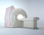
Device Information and Specifi cation
CLINICAL APPLICATION
Whole body
CONFIGURATION
Cylindrical Wide Short Bore
Opt. (WIP) Single and Multi Voxel
SE, FE, IR, FastSE, FastIR, FastFLAIR, Fast STIR, FastFE, FASE, Hybrid EPI, Multi Shot EPI; Angiography: 2D(gate/non-gate)/3D TOF, SORS-STC
IMAGING MODES
Single, multislice, volume study
TE
8 msec min. SE; 1.2 msec min. FE
less than 0.015 (256x256)
1.0 min. 2-DFT: 0.2 min. 3-DFT
32-1024, phase;; 64-1024, freq.
65.5 cm, patient aperture
4050 kg (bare magnet incl. L-He)
COOLING SYSTEM TYPE
Closed-loop water-cooled
Liquid helium: approx. less than 0.05 L/hr
Passive, active, auto-active
| | | | | |
| | | | | |
| |
|
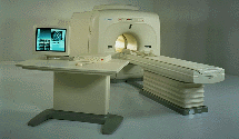
Device Information and Specifi cation
CLINICAL APPLICATION
Whole body
CONFIGURATION
Cylindrical Wide Short Bore
SE, FE, IR, FastSE, FastIR, FastFLAIR, Fast STIR, FastFE, FASE, Hybrid EPI, Multi Shot EPI; Angiography: 2D(gate/non-gate)/3D TOF, SORS-STC
IMAGING MODES
Single, multislice, volume study
TE
8 msec min. SE; 0.9 msec min. FE
less than 0.011 (256x256)
1.0 min. 2-DFT: 0.2 min. 3-DFT
32-1024, phase;; 64-1024, freq.
65.5 cm, patient aperture
4050 kg (bare magnet incl. L-He)
POWER REQUIREMENTS
380/400/415/440/480 V
COOLING SYSTEM TYPE
Closed-loop water-cooled
Liquid helium: approx. less than 0.05 L/hr
Passive, active, auto-active
| | | |
• View the DATABASE results for 'Excelart XG™ with Pianissimo' (2).
| | | | |  Further Reading: Further Reading: | News & More:
|
|
| |
| | | | | |
| |
|
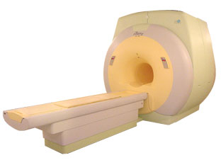
From Toshiba America Medical Systems Inc.;
FLEXART™ series is a 0.5 T superconducting MRI system that has been designed to meet the expanding role of MRI in today's clinical environment. The system utilizes innovative technologies such as digital RF, high speed actively shielded gradients and optimized RF coils which support a wide range of MRI developments.
Device Information and Specifi cation
CLINICAL APPLICATION
Whole body
Quadrature, solenoid and multi-channel configurations
SE, FE, IR, FastSE, FastIR, FastFLAIR, Fast STIR, FastFE, FASE, Hybrid EPI, Multi Shot EPI; Angiography: 2D(gate/non-gate)/3D TOF, SORS-STC
IMAGING MODES
Single, multislice, volume study
POWER REQUIREMENTS
380/400/415/440/480 V
COOLING SYSTEM TYPE
Closed-loop water-cooled
| | | |
• View the DATABASE results for 'FLEXART™' (2).
| | | | |
| | | Searchterm 'Cation' was also found in the following services: | | | | |
| | |
| |
|
From ISOL Technology
'Ultra high field MR system, it's right close to you.
FORTE 3.0T is the new standard for the future ultra high field MR system.
If you are pushing the limits of your existing clinical MR scanner, the FORTE will surely take you to the next level of diagnostic imaging.
FORTE is the core leader of the medical technology in the 21st century. Proving effects of fMRI that cannot be measured with MRI less than 2.0T.'
Device Information and Specifi cation
CLINICAL APPLICATION
Whole body
CONFIGURATION
Short bore compact
128 x 128, 256 x 256, 512 x 512, 1024 x 1024
| | | |
• View the DATABASE results for 'FORTE 3.0T™' (2).
| | | | |
| | | | | |
| |
|
In electromagnetism, the Faraday cage or shield is an appli cation of Gauss's law, one of Maxwell's equations. Gauss's law describes the distribution of electrical charge on a conducting form, such as a sphere, a plane, a torus, etc. Intuitively, since like charges repel each other, charge will "migrate" to the surface of the conducting form, as described below. The appli cation is named after physicist Michael Faraday, who built the first Faraday cage in 1836, to demonstrate his finding. A Faraday shield is used generally for any kind of electrostatic shielding.
In MRI, one use of the Faraday shield is the shielding of the scanning room, to block incoming radio frequency (RF) signals which would contaminate the send and received signals of the MRI scanner, and it suppresses RF signals, which would else pollute the environment around. | | | |
• View the DATABASE results for 'Faraday Shield' (5).
| | | | | | Further Reading: | | Basics:
|
|
News & More:
| |
| |
| | | | |
| | |
| | | |
|
| |
 | Look
Ups |
| |