 | Info
Sheets |
| | | | | | | | | | | | | | | | | | | | | | | | |
 | Out-
side |
| | | | |
|
| | | | | |  | Searchterm 'Cation' was also found in the following services: | | | | |
|  | |  | |  |  |  |
| |
|

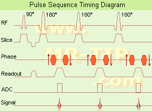
(FSE) In the pulse sequence timing diagram, a fast spin echo sequence with an echo train length of 3 is illustrated.
This sequence is characterized by a series of rapidly applied 180° rephasing pulses and multiple echoes, changing the phase encoding gradient for each echo.
The echo time TE may vary from echo to echo in the echo train. The echoes in the center of the K-space (in the case of linear k-space acquisition) mainly produce the type of image contrast, whereas the periphery of K-space determines the spatial resolution. For example, in the middle of K-space the late echoes of T2 weighted images are encoded. T1 or PD contrast is produced from the early echoes.
The benefit of this technique is that the scan duration with, e.g. a turbo spin echo turbo factor / echo train length of 9, is one ninth of the time. In T1 weighted and proton density weighted sequences, there is a limit to how large the ETL can be (e.g. a usual ETL for T1 weighted images is between 3 and 7). The use of large echo train lengths with short TE results in blurring and loss of contrast. For this reason, T2 weighted imaging profits most from this technique.
In T2 weighted FSE images, both water and fat are hyperintense. This is because the succession of 180° RF pulses reduces the spin spin interactions in fat and increases its T2 decay time. Fast spin echo (FSE) sequences have replaced conventional T2 weighted spin echo sequences for most clinical appli cations. Fast spin echo allows reduced acquisition times and enables T2 weighted breath hold imaging, e.g. for appli cations in the upper abdomen.
In case of the acquisition of 2 echoes this type of a sequence is named double fast spin echo / dual echo sequence, the first echo is usually density and the second echo is T2 weighted image. Fast spin echo images are more T2 weighted, which makes it difficult to obtain true proton density weighted images. For dual echo imaging with density weighting, the TR should be kept between 2000 - 2400 msec with a short ETL (e.g., 4).
Other terms for this technique are:
Turbo Spin Echo
Rapid Imaging Spin Echo,
Rapid Spin Echo,
Rapid Acquisition Spin Echo,
Rapid Acquisition with Refocused Echoes
| | | |  | |
• View the DATABASE results for 'Fast Spin Echo' (31).
| | | | |  Further Reading: Further Reading: | | Basics:
|
|
News & More:
| |
| |
|  | |  |  |  |
| |
|
A brand name for ferumoxide (same as Endorem™)
Feridex® is a sterile aqueous colloid of superparamagnetic iron oxide associated with dextran for intravenous administration as a MRI contrast medium for the detection of liver lesions that are associated with an alteration in the RES.
Feridex® is taken up by macrophages, found only in healthy liver cells but not in most tumors. Tissues such as metastases, primary liver cancer, cysts and various benign tumors, adenomas and hyperplasia retain their native signal intensity, so the contrast between normal and abnormal tissue is increased.
Feridex® is a black to reddish-brown aqueous colloid.
See also Ferumoxide. In November 2008, AMAG Pharmaceuticals, Inc. decided to discontinue the manufacturing of Feridex.
Drug Information and Specification
T2, predominantly negative enhancement
r1=40.0, r2=160, B0=0.47T
PHARMACOKINETIC
RES-directed
CONCENTRATION
11.2mg Fe/ml
PREPARATION
Suspend in an isotonic glucose solution
DEVELOPMENT STAGE
For sale
PRESENTATION
Ampoule of 8 mL
DO NOT RELY ON THE INFORMATION PROVIDED HERE, THEY ARE
NOT A SUBSTITUTE FOR THE ACCOMPANYING
PACKAGE INSERT!
Distribution Information
TERRITORY
TRADE NAME
DEVELOPMENT
STAGE
DISTRIBUTOR
| |  | |
• View the DATABASE results for 'Feridex®' (9).
| | | | |  Further Reading: Further Reading: | Basics:
|
|
News & More:
| |
| |
|  |  | Searchterm 'Cation' was also found in the following services: | | | | |
|  |  |
| |
|
A solution of ferric ammonium citrate (Geritol) used to enhance the delineation of the bowel. With T1 weighted magnetic resonance imaging ( MRI) the predominantly positive enhancement helps to distinguish organs and tissues that are adjacent to the upper regions of the gastrointestinal tract. Product name found as both Ferriseltz® and FerriSeltz®.
Drug Information and Specification
T1, Predominantly positive enhancement
PHARMACOKINETIC
Gastrointestinal
DEVELOPMENT STAGE
For sale
PRESENTATION
Bags with powder
DO NOT RELY ON THE INFORMATION PROVIDED HERE, THEY ARE
NOT A SUBSTITUTE FOR THE ACCOMPANYING
PACKAGE INSERT!
Distribution Information
TERRITORY
TRADE NAME
DEVELOPMENT
STAGE
DISTRIBUTOR
USA
FerriSeltz®
for sale
| |  | |
• View the DATABASE results for 'FerriSeltz®' (4).
| | | | |
|  | |  |  |  |
| |
|
Flow phenomena are intrinsic processes in the human body. Organs like the heart, the brain or the kidneys need large amounts of blood and the blood flow varies depending on their degree of activity. Magnetic resonance imaging has a high sensitivity to flow and offers accurate, reproducible, and noninvasive methods for the quantifi cation of flow. MRI flow measurements yield information of blood supply of of various vessels and tissues as well as cerebro spinal fluid movement.
Flow can be measured and visualized with different pulse sequences (e.g. phase contrast sequence, cine sequence, time of flight angiography) or contrast enhanced MRI methods (e.g. perfusion imaging, arterial spin labeling).
The blood volume per time (flow) is measured in: cm3/s or ml/min. The blood flow-velocity decreases gradually dependent on the vessel diameter, from approximately 50 cm per second in arteries with a diameter of around 6 mm like the carotids, to 0.3 cm per second in the small arterioles.
Different flow types in human body:
•
Behaves like stationary tissue, the signal intensity depends on T1, T2 and PD = Stagnant flow
•
Flow with consistent velocities across a vessel = Laminar flow
•
Laminar flow passes through a stricture or stenosis (in the center fast flow, near the walls the flow spirals) = Vortex flow
•
Flow at different velocities that fluctuates = Turbulent flow
See also Flow Effects, Flow Artifact, Flow Quantification, Flow Related Enhancement, Flow Encoding, Flow Void, Cerebro Spinal Fluid Pulsation Artifact, Cardiovascular Imaging and Cardiac MRI. | | | |  | |
• View the DATABASE results for 'Flow' (113).
| | |
• View the NEWS results for 'Flow' (7).
| | | | |  Further Reading: Further Reading: | News & More:
|
|
| |
|  | |  |  |
|  | | |
|
| |
 | Look
Ups |
| |