 | Info
Sheets |
| | | | | | | | | | | | | | | | | | | | | | | | |
 | Out-
side |
| | | | |
|
| | | | | |  | Searchterm 'Cation' was also found in the following services: | | | | |
|  |  |
| |
|
(DICOM) DICOM is the industry standard for transferral of radiologic images and other medical information between computers. Patterned after the Open System Interconnection of the International Standards Organization, DICOM enables digital communi cation between diagnostic and therapeutic equipment and systems from various manufacturers.
The DICOM 3.0 standard evolved from versions 1.0 (1985) and 2.0 (1988) of a standard developed by the American College of Radiology (ACR) and National Electrical Manufacturers Association (NEMA). To support the implementation and demonstration of DICOM 3.0, the RSNA Electronic Communi cations Committee began to work with the ACR-NEMA MedPacs ad hoc section in 1992.
Also Picture Archiving and Communication Systems (PACS), which are connected with the Radiology Information System (RIS) use commonly the DICOM standard for the transfer and storage of medical images. | | | | | |  Further Reading: Further Reading: | | Basics:
|
|
News & More:
| |
| |
| | | | |  |
| |
|

Swiss-based, formerly Bruker AG - split on the 5th October 2001 into the groups: Bruker Daltonics ( Mass spectroscopy), Bruker Optics (Infrared spectroscopy), Bruker AXS (X-ray spectroscopy) and Bruker BioSpin (The largest part, the NMR business core, the EPR and the Tomography activities).
Product Lines:
•
PharmaScan® - MRI//MRS systems tailored to high-throughput and routine appli cations in pharmaceutical research.
Product Specification
Contact Information
Please see Bruker BioSpin AG's
| | | |
• View the NEWS results for 'Bruker BioSpin AG' (1).
| | | | | | Further Reading: | News & More:
|
|
| |
| | | | | |
| |
|
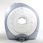
From GE Healthcare;
GE Healthcare has added the Signa HDe 1.5T™, a compact MRI device at an affordable price to its family of MRI products. It has a single electronic cabinet that can be positioned inside the scanner room rather than
in a separate equipment room. The Signa HDe 1.5T can be installed in the same physical lo cation as 0.5T MRI systems with minimal construction costs. According to GE, the installation has been simplified to last only 7 days and has a 30 percent smaller footprint than a typical 1.5T system.
The 1.5T Signa™ HDe MRI system is substantially equivalent to the currently marketed GE 1.5T machines. The data acquisition system supports 1, 4, 8 independent receive channels and multiple independent coil elements per channel during a single acquisition series. The gradient specifi cations of HDe are lower than other GE Signa 1.5T MRI systems, but it can support clinical appli cations in cardiac and spectroscopy imaging.
Device Information and Specifi cation CLINICAL APPLICATION Whole body CONFIGURATION Compact short bore 2D 0.7 mm to 20 mm; 3D 0.1 mm to 5 mm 128x512 steps 32 phase encode POWER REQUIREMENTS 480 or 380/415 less than 0.03 L/hr liquid helium | | | |
• View the NEWS results for 'Signa HDe 1.5T™' (1).
| | | | | | Further Reading: | Basics:
|
|
| |
| | | Searchterm 'Cation' was also found in the following services: | | | | |
| | |
| |
|
Sinerem® is the brand name (same as Combidex®) for an ultrasmall superparamagnetic iron oxide ( USPIO) to detect metastatic disease in lymph nodes. Metastatic nodes show less uptake of this MRI contrast agent, which results in less signal decrease and allows the differentiation of normal lymph nodes from normal-sized, metastatic nodes.
Lymph node imaging with Sinerem® is performed 24 to 36 hours after slow infusion. Normal lymph nodes turn black post contrast, namely on T2* weighted images. Metastatic lymph nodes remain unchanged in signal intensity.
Indi cation and Diseases: Cancer, Imaging for diagnosis, Lymphatic disorders.
See Ferumoxtran, and Classifications, Characteristics, etc.
Guerbet decided in 2007 to withdraw its Marketing Authorisation Application
(MAA) for Sinerem.Drug Information and Specification r1=25, r2=160, B0=0.47T, r1=23.3, r2=48.9, B0=0.47T PHARMACOKINETIC Vascular, lymph v. hepatocyte (AG-USPIO) PREPARATION Suspend in an isotonic glucose solution DO NOT RELY ON THE INFORMATION PROVIDED HERE, THEY ARE
NOT A SUBSTITUTE FOR THE ACCOMPANYING PACKAGE INSERT! | | | |
• View the DATABASE results for 'Sinerem®' (6).
| | | | | | Further Reading: | Basics:
|
|
News & More:
| |
| |
| | | | | |
| |
|
If the receiving RF coil is sensitive to tissue signal arising from outside the desired FOV, this undesired signal may be incorrectly mapped to a lo cation within the image, a phenomenon known as aliasing. This is a consequence of the acquired k-space frequencies not being sampled densely enough, whereby portions of the object outside of the desired FOV get mapped to an incorrect lo cation inside the FOV.
The sampling frequency should be at least twice the frequency being sampled. The maximum measurable frequency is therefore equal to half the sampling frequency. This is the so-called Nyquist limit. When the frequency is higher than the Nyquist limit, aliasing occurs.
A similar problem occurs in the phase encoding direction, where the phases of signal-bearing tissues outside of the FOV in the y-direction are a repli cation of the phases that are encoded within the FOV. This signal will be mapped, or wrapped back into the image at incorrect lo cations, and is seen as artifact.
See also Aliasing Artifact. | | | |
• View the DATABASE results for 'Aliasing' (19).
| | | | | | Further Reading: | News & More:
|
|
| |
| | | | |
| | | |
|
| |
 | Look
Ups |
| |


