| Info
Sheets |
| | | | | | | | | | | | | | | | | | | | | | | | |
 | Out-
side |
| | | | |
|
| | | | | |  | Searchterm 'Cation' was also found in the following services: | | | | |
|  |  |
| |
|
The characteristics of a hepatobiliary contrast agent are specific liver uptake and excretion via the biliary system. The paramagnetic substance (e.g. manganese, gadolinium) is taken up by normal hepatocytes. Diseased liver tissue did not include hepatocytes or their function is disturbed. Therefore, the signal of healthy liver tissue increases on T1 weighted sequences, but not in the liver lesions.
Another type of liver imaging contrast agent is superparamagnetic iron oxide. These particles accumulate in the reticuloendothelial system (RES) of the liver, and darken the healthy liver tissue in T2 weighted images. RES cells (including Kupffer cells) are existing in healthy liver tissue, in altered tissue with reduced RES activity or without RES cells the contrast agent concentration is also low or not existing, which improves the liver to lesion contrast.
Benefits of hepatobiliary contrast agents:
•
Liver lesions (e.g., tumor, metastases, haemangioma etc.) are better detectable and to characterize.
•
These contrast agents are useful to analyze and evaluate the liver function (in cases of diffuse liver diseases e.g., cirrhosis).
•
Imaging of the gallbladder and biliary system is improved.
Differences of a hepatobiliary contrast agent compared with a targeted contrast agent for Kupffer cells:
•
The higher number of hepatocytes than Kupffer cells improves the uptake effectiveness of the contrast agent.
•
Hepatobiliary contrast agents enable a better opacifi cation of the biliary ducts and the gallbladder caused by the biliary excretion.
•
Hepatobiliary contrast media are fast excreted agents. RES targeted contrast agents remain longer in the body, a fact that can increase possible side effects.
See also Superparamagnetic Contrast Agents, Hepatobiliary Chelates, Liver Imaging, Endoremâ„¢, Primovistâ„¢, and Classifications, Characteristics, etc.
See also the related poll result: ' The development of contrast agents in MRI is' | | | | | |  Further Reading: Further Reading: | | Basics:
|
|
News & More:
| |
| |
| | | | | |
| |
|
| | | |
• View the DATABASE results for 'Imaging Coil' (7).
| | |
• View the NEWS results for 'Imaging Coil' (9).
| | | | | | Further Reading: | Basics:
|
|
| |
| | | | | |
| |
|
Knee and shoulder MRI exams are the most commonly requested musculoskeletal MRI scans. Other MR imaging of the extremities includes hips, ankles, elbows, and wrists. Orthopedic imaging requires very high spatial resolution for reliable small structure definition and therefore places extremely high demands on SNR.
Exact presentation of joint pathology expects robust and reliable fat suppression, often under difficult conditions like off-center FOV,
imaging at the edge of the field homogeneity or in regions with complex magnetic susceptibility.
MR examinations can evaluate meniscal dislo cations, muscle fiber tears, tendon disruptions, tendinitis, and diagnose bone tumors and soft tissue masses. MR can also demonstrate acute fractures that are radiographically impossible to see. Evaluation of articular cartilage for traumatic injury or assessment of degenerative disease represents an imaging challenge, which can be overcome by high field MRI appli cations. Currently, fat-suppressed 3D spoiled gradient echo sequences and density weighted fast spin echo sequences are the gold-standard techniques used to assess articular cartilage.
Open MRI procedures allow the kinematic imaging of joints, which provides added value to any musculoskeletal MRI practice. This technique demonstrates the actual functional impingements or positional subluxations of joints. In knee MRI examinations, the kinematical patellar study can show patellofemoral joint abnormalities.
See also Open MRI, Knee MRI, Low Field MRI. | | | | | | | | | | |
• View the DATABASE results for 'Imaging of the Extremities' (5).
| | | | | | Further Reading: | Basics:
|
|
News & More:
| |
| |
| | | Searchterm 'Cation' was also found in the following services: | | | | |
| | |
| |
|
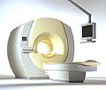
From Philips Medical Systems;
Philips Infinion 1.5 T is designed to maximize the efficiency and quality of patient care. Developed with the patient in mind, the Infinion is the shortest and most open 1.5T scanner available. The unique 'ultra short' 1.4 m magnet assures patient comfort and acceptance without compromising image quality and clinical performance.
Device Information and Specifi cation
CLINICAL APPLICATION
Whole body
CONFIGURATION
Ultra short bore
Head, head / neck, integrated C-spine, L/T spine array, small large GP coils, body flex array, torso pelvis array, breast array, endocavitary, shoulder array, lower extremity, hand / wrist, cardiac, PV array
SE, TSE, SS TSE, EPI, IR, STIR, FLAIR, FFE, TFE, T1 TFE, T2 TFE, Presat, Fatsat, MTC, Diff-opt., Angiography: PCA, MCA, TOF
IMAGING MODES
Single slice, single volume, multi slice, multi volume
80 images/sec std.; up to320 opt.@256
H*W*D
233 (lead fitted) x 198 x 140 cm
POWER REQUIREMENTS
400/480 V
COOLING SYSTEM TYPE
Closed loop, chilled water
| | | |
• View the DATABASE results for 'Infinion 1.5T™' (2).
| | | | |
| | | | | |
| |
|
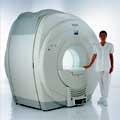
Device Information and Specifi cation
CLINICAL APPLICATION
Whole body
CONFIGURATION
Short bore compact
Standard: head, body, C1, C3; Optional: Small joint, flex-E, flex-R, endocavitary (L and S), dual TMJ, knee, neck, T/L spine, breast; Optional phased array: Spine, pediatric, 3rd party connector, Flex-S-M-L, flex body, flex cardiac, neuro-vascular, head
SE, Modified-SE ( TSE), DAVE, STIR, FLAIR, SPIR, MTC, Dynamic, Keyhole, CLEAR, Q Flow, Balanced FFE, Multi Chunk 3D, Multi Stack 3D, FFE-EPI, SE-EPI, IR-EPI, GRASE, Diffusion Imaging, Perfusion Imaging;; Angiography: Inflow MRA, TONE, PCA, CE MRA
RapidView Recon. greater than 500 @ 256 Matrix
128 x 128, 256 x 256,512 x 512,1024 x 1024
Variable in 1% increments
Lum.: 120 cd/m2; contrast: 150:1
Variable (op. param. depend.)
POWER REQUIREMENTS
380/400 V
| | | |
• View the DATABASE results for 'Intera 0.5T™' (2).
| | | | |
| | | | |
| | | |
|
| |
 | Look
Ups |
| |