 | Info
Sheets |
| | | | | | | | | | | | | | | | | | | | | | | | |
 | Out-
side |
| | | | |
|
| | | | | |  | Searchterm 'Cation' was also found in the following services: | | | | |
|  |  |
| |
|

The company is a leading manufacturer and developer of magnetic resonance imaging ( MRI) scanners.
The Patient Friendly MRI Company, formed in 1978, is engaged in the business of inventing, manufacturing, selling and servicing magnetic resonance imaging ( MRI) scanners. FONAR is the oldest MRI company in the world. After receiving hundreds of millions in a windfall from protecting their MRI patents, they made a MRI scanner that no other MRI manufacturer has. One that the patient stands in and they call Indomitable, the Stand-Up MRI. Patients like it because it is the least claustrophobic, most comfortable MRI on the market. Doctors like it because of its superior image quality and for the first time, the patient can be scanned in the weight-bearing position, or the position of pain or symptom. In October of 2004, the company changed the product name of the Stand-Up MRI to the Upright MRI. Fonar introduced the first "open" MRI scanner in 1980 and is the originator of the iron-core nonsuperconductive and permanent magnet technology.
MRI Scanners:
- 0.6T:
•
QUAD™ 12000 - Its 19-inch gap and Whisper Gradients™ make it extraordinarily spacious, quiet and comfortable. With its signal to noise advantage of 0.6 T and its comprehensive array of Organ-Specific™ receiver coils, the QUAD™ 12000 provides high-speed, high resolution and high contrast scanning.
Product Specification
•
OR 360°™ - cleared for marketing by the FDA in March 2000, 360° access to the patient. A dual-purpose scanner, it can be used for conventional diagnostic scanning when not in surgical mode.
Product Specification
Contact Information
MAIL
FONAR Corporation
110 Marcus Drive
Melville, N.Y. 11747
USA
| | | |
• View the NEWS results for 'FONAR Corporation' (87).
| | | | |  Further Reading: Further Reading: | | Basics:
|
|
News & More:
| |
| |
| | | | | |
| |
|
The FEER method was the first clinically useful flow quantification method using phase effects, from which all spin phase related flow quantification techniques currently in use are derived.
In this sequence a gradient echo is measured after a gradient with flow compensation. The measured signal phase should be zero for all pixels. A deviation from gradient symmetry by shifting the gradient ramp slightly away from the symmetry condition will impart a defined phase shift to the magnetization vectors associated with spins from pixels with flow.
Slight stable variations in the magnetic field across the imaging volume will prevent the phase angle from being uniformly zero throughout the volume in the flow-compensated image.
The first image (acquired without gradient shift) serves as reference, defining the values of all pixel phase angles in the flow (motion) compensated sequence.
Ensuing images with gradient phase shifts imparted in each of the 3 spatial axes will then permit measurement of the 3 components of the velocity vector v = (vx, vy, vz) by calculating the respective phases px, py and pz by simply subtracting the pixel phases measured in the compensated image from the 3 images with a well defined velocity sensitization.
The determination of all 3 components of the velocity vector requires the measurement of 4 images.
The phase quantifi cation requires an imaging time four times longer than the simple measurement of a phase image and associated magnitude image.
If only one arbitrary flow direction is of interest, it suffices to acquire the reference image plus one image velocity sensitized in the arbitrary direction of interest.
See also Flow Quantification. | | | | | |
| | | | | |
| |
|
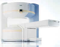
From Siemens Medical Systems;
the MAGNETOM Rhapsody™. This open MRI system offers the proven image
quality of 1.0 Tesla. In addition to the resulting broad range of appli cations, the open magnet of the high field system MAGNETOM Rhapsodyâ„¢ facilitates examination of claustrophobic and pediatric patients. And the system allows for expanded interventional appli cations.
Device Information and Specifi cation CLINICAL APPLICATION Whole body GRE, IR, FIR, STIR, TrueIR/FISP, FSE, FLAIR, MT, SS-FSE, MT-SE, MTC, MSE, EPI, GMR, fat/water sat./exc. IMAGING MODES Single, multislice, volume study, multi angle, multi oblique1024 x 1024 full screen display POWER REQUIREMENTS 380/400/420/440/480 V | | | |
• View the DATABASE results for 'MAGNETOM Rhapsody™' (2).
| | | | |
| | | Searchterm 'Cation' was also found in the following services: | | | | |
| | |
| |
|

It is important to remember when working around a superconducting magnet that the magnetic field is always on. Under usual working conditions the field is never turned off. Attention must be paid to keep all ferromagnetic items at an adequate distance from the magnet. Ferromagnetic objects which came accidentally under the influence of these strong magnets can injure or kill individuals in or nearby the magnet, or can seriously damage every hardware, the magnet itself, the cooling system, etc..
See MRI resources Accidents.
The doors leading to a magnet room should be closed at all times except when entering or exiting the room. Every person working in or entering the magnet room or adjacent rooms with a magnetic field has to be instructed about the dangers. This should include the patient, intensive-care staff, and maintenance-, service- and cleaning personnel, etc..
The 5 Gauss limit defines the 'safe' level of static magnetic field exposure. The value of the absorbed dose is fixed by the authorities to avoid heating of the patient's tissue and is defined by the specific absorption rate.
Leads or wires that are used in the magnet bore during imaging procedures, should not form large-radius wire loops. Leg-to-leg and leg-to-arm skin contact should be prevented in order to avoid the risk of burning due to the generation of high current loops if the legs or arms are allowed to touch. The patient's skin should not be in contact with the inner bore of the magnet.
The outflow from cryogens like liquid helium is improbable during normal operation and not a real danger for patients.
The safety of MRI contrast agents is tested in drug trials and they have a high compatibility with very few side effects. The variations of the side effects and possible contraindications are similar to X-ray contrast medium, but very rare. In general, an adverse reaction increases with the quantity of the MRI contrast medium and also with the osmolarity of the compound.
See also 5 Gauss Fringe Field, 5 Gauss Line, Cardiac Risks, Cardiac Stent, dB/dt, Legal Requirements, Low Field MRI, Magnetohydrodynamic Effect, MR Compatibility, MR Guided Interventions, Claustrophobia, MRI Risks and Shielding. | | | | | | | | |
• View the DATABASE results for 'MRI Safety' (42).
| | |
• View the NEWS results for 'MRI Safety' (13).
| | | | | | Further Reading: | | Basics:
|
|
News & More:
| |
| |
| | | | | |
| |
|
The definition of imaging is the visual representation of an object. Medical imaging began after the discovery of x-rays by Konrad Roentgen 1896. The first fifty years of radiological imaging, pictures have been created by focusing x-rays on the examined body part and direct depiction onto a single piece of film inside a special cassette. The next development involved the use of fluorescent screens and special glasses to see x-ray images in real time.
A major development was the appli cation of contrast agents for a better image contrast and organ visualization. In the 1950s, first nuclear medicine studies showed the up-take of very low-level radioactive chemicals in organs, using special gamma cameras. This medical imaging technology allows information of biologic processes in vivo. Today, PET and SPECT play an important role in both clinical research and diagnosis of biochemical and physiologic processes. In 1955, the first x-ray image intensifier allowed the pick up and display of x-ray movies.
In the 1960s, the principals of sonar were applied to diagnostic imaging. Ultrasonic waves generated by a quartz crystal are reflected at the interfaces between different tissues, received by the ultrasound machine, and turned into pictures with the use of computers and reconstruction software. Ultrasound imaging is an important diagnostic tool, and there are great opportunities for its further development. Looking into the
future, the grand challenges include targeted contrast agents, real-time 3D ultrasound imaging, and molecular imaging.
Digital imaging techniques were implemented in the 1970s into conventional fluoroscopic image intensifier and by Godfrey Hounsfield with the first computed tomography. Digital images are electronic snapshots sampled and mapped as a grid of dots or pixels. The introduction of x-ray CT revolutionised medical imaging with cross sectional images of the human body and high contrast between different types of soft tissue. These developments were made possible by analog to digital converters and computers. The multislice spiral CT technology has expands the clinical appli cations dramatically.
The first MRI devices were tested on clinical patients in 1980. The spread of CT machines is the spur to the rapid development of MRI imaging and the introduction of tomographic imaging techniques into diagnostic nuclear medicine. With technological improvements including higher field strength, more open MRI magnets, faster gradient systems, and novel data-acquisition techniques, MRI is a real-time interactive imaging modality that provides both detailed structural and functional information of the body.
Today, imaging in medicine has advanced to a stage that was inconceivable 100 years ago, with growing medical imaging modalities:
•
Single photon emission computed tomography (SPECT)
•
Positron emission tomography (PET)
All this type of scans are an integral part of modern healthcare.
Because of the rapid development of digital imaging modalities, the increasing need for an efficient management leads to the widening of radiology information systems (RIS) and archival of images in digital form in picture archiving and communication systems (PACS).
In telemedicine, healthcare professionals are linked over a computer network. Using cutting-edge computing and communi cations technologies, in videoconferences, where audio and visual images are transmitted in real time, medical images of MRI scans, x-ray examinations, CT scans and other pictures are shareable.
See also Hybrid Imaging.
See also the related poll results: ' In 2010 your scanner will probably work with a field strength of', ' MRI will have replaced 50% of x-ray exams by' | | | | | | | | |
• View the DATABASE results for 'Medical Imaging' (20).
| | |
• View the NEWS results for 'Medical Imaging' (81).
| | | | | | Further Reading: | Basics:
|
|
News & More:
| |
| |
| | | | |
| | | |
|
| |
 | Look
Ups |
| |


