 | Info
Sheets |
| | | | | | | | | | | | | | | | | | | | | | | | |
 | Out-
side |
| | | | |
|
| | | | | |  | Searchterm 'Cation' was also found in the following services: | | | | |
|  |  |
| |
|
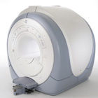
From GE Healthcare;
The Signa HDx MRI system is GE's leading edge whole body magnetic resonance scanner designed to support high resolution, high signal to noise ratio, and short scan times.
Signa HDx 3.0T offers new technologies like ultra-fast image reconstruction through the new XVRE recon engine, advancements in parallel imaging algorithms and the broadest range of premium appli cations. The HD appli cations, PROPELLER (high-quality brain imaging extremely resistant to motion artifacts), TRICKS (contrast-enhanced angiographic vascular lower leg imaging), VIBRANT (for breast MRI), LAVA (high resolution liver imaging with shorter breath holds and better organ coverage) and MR Echo (high-definition cardiac images in real time) offer unique capabilities.
Device Information and Specifi cation CLINICAL APPLICATION Whole body
CONFIGURATION Compact short bore SE, IR, 2D/3D GRE, RF-spoiled GRE, 2DFGRE, 2DFSPGR, 3DFGRE, 3DFSPGR, 3DTOFGRE, 3DFSPGR, 2DFSE, 2DFSE-XL, 2DFSE-IR, T1-FLAIR, SSFSE, EPI, DW-EPI, BRAVO, Angiography: 2D/3D TOF, 2D/3D phase contrast vascular IMAGING MODES Single, multislice, volume study, fast scan, multi slab, cine, localizer H*W*D 240 x 2216,6 x 201,6 cm POWER REQUIREMENTS 480 or 380/415, 3 phase ||
COOLING SYSTEM TYPE Closed-loop water-cooled grad. | | | | | |
| | | | | |
| |
|
Quick Overview
Please note that there are different common names for this MRI artifact.
DESCRIPTION
Image wrap around
Aliasing is an artifact that occurs in MR images when the scanned body part is larger than field of view ( FOV). As a consequence of the acquired k-space frequencies not being sampled densely enough, whereby portions of the object outside of the desired FOV get mapped to an incorrect lo cation inside the FOV. The cyclical property of the Fourier transform fills the missing data of the right side with data from behind the FOV of the left side and vice versa. This is caused by a too small number of samples acquired in, e.g. the frequency encoding direction, therefore the spectrums will overlap, resulting in a repli cation of the object in the x direction.
Aliasing in the frequency direction can be eliminated by twice as fast sampling of the signal or by applying frequency specific filters to the received signal.
A similar problem occurs in the phase encoding direction, where the phases of signal-bearing tissues outside of the FOV in the y-direction are a repli cation of the phases that are encoded within the FOV. Phase encoding gradients are scaled for the field of view only, therefore tissues outside the FOV do not get properly phase encoded relative to their actual position and 'wraps' into the opposite side of the image.

Image Guidance
| | | |
• View the DATABASE results for 'Aliasing Artifact' (11).
| | | | |
| | | | | |
| |
|
Drug Information and Specification
r1=25, r2=160, B0=0.47T, r1=23.3, r2=48.9, B0=0.47T
PHARMACOKINETIC
Vascular, lymph v. hepatocyte (AG-USPIO)
PREPARATION
Suspend in an isotonic glucose solution
DO NOT RELY ON THE INFORMATION PROVIDED HERE, THEY ARE
NOT A SUBSTITUTE FOR THE ACCOMPANYING
PACKAGE INSERT!
| | | |
• View the DATABASE results for 'Combidex®' (6).
| | |
• View the NEWS results for 'Combidex®' (1).
| | | | |  Further Reading: Further Reading: | | Basics:
|
|
News & More:
| |
| |
| | | Searchterm 'Cation' was also found in the following services: | | | | |
| | |
| |
|
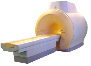
'MRI system is not an expensive equipment anymore.
ENCORE developed by ISOL Technology is a low cost MRI system with the advantages like of the 1.0T MRI scanner. Developed specially for the overseas market, the ENCORE is gaining popularity in the domestic market by medium sized hospitals.
Due to the optimum RF and Gradient appli cation technology. ENCORE enables to obtain high resolution imaging and 2D/3D Angio images which was only possible in high field MR systems.'
- Less consumption of the helium gas due to the ultra-lightweight magnet specially designed and manufactured for ISOL.
- Cost efficiency MR system due to air cooling type (equivalent to permanent magnetic).
- Patient processing speed of less than 20 minutes.'
Device Information and Specifi cation
CLINICAL APPLICATION
Whole body
CONFIGURATION
Short bore compact
| | | |
• View the DATABASE results for 'ENCORE 0.5T™' (2).
| | | | |
| | | | | |
| |
|
A problem occurs in the phase encoding direction, where the phases of signal-bearing tissues outside of the FOV in the y-direction are a repli cation of the phases that are encoded within the FOV. This signal will be mapped (wrapped, backfolded) back into the image at incorrect lo cations.
Foldover suppression ( phase oversampling, no phase wrap) is a user-selectable parameter that maps this signal to its correct lo cation outside the FOV, then discards any signal from outside the FOV before displaying the image. In order to be able to choose this parameter, in most cases more than an average is necessary.
See also Phase Wrapping Artifact and Oversampling. | | | |
• View the DATABASE results for 'Foldover Suppression' (4).
| | | | |
| | | | |
| | |
| | | |
|
| |
 | Look
Ups |
| |


