| Info
Sheets |
| | | | | | | | | | | | | | | | | | | | | | | | |
 | Out-
side |
| | | | |
|
| | | | | |  | Searchterm 'Cation' was also found in the following services: | | | | |
|  |  |
| |
|
Device Information and Specifi cation CLINICAL APPLICATION Whole body Quadrature, solenoid and multi-channel configurations SE, FE, IR, FastSE, FastIR, FastFLAIR, Fast STIR, FastFE, FASE, Hybrid EPI, Multi Shot EPI; Angiography: 2D(gate/non-gate)/3D TOF, SORS-STC IMAGING MODES Single, multislice, volume study POWER REQUIREMENTS 380/400/415/440/480 V COOLING SYSTEM TYPE Cryogenless | | | | | |
| | | | | |
| |
|
Drug Information and Specification T1, Predominantly positive enhancement PHARMACOKINETIC Intravascular, extracellular, renal excretion CONCENTRATION 287 mg/mL,0.5 mol/L DOSAGE 0.1-0.2 mmol/kg / 0.2-0.4 ml/kg PREPARATION Finished product INDICATION Neuro/whole body DEVELOPMENT STAGE For sale PRESENTATION Vials of 10 mL, 15 mL and 20 mL
DO NOT RELY ON THE INFORMATION PROVIDED HERE, THEY ARE
NOT A SUBSTITUTE FOR THE ACCOMPANYING PACKAGE INSERT! Distribution Information TERRITORY TRADE NAME DEVELOPMENT
STAGE DISTRIBUTOR Australia Omniscan® for sale | | | |
• View the DATABASE results for 'Omniscan®' (7).
| | | | |  Further Reading: Further Reading: | | Basics:
|
|
News & More:
| |
| |
| | | | | |
| |
|
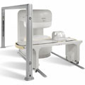
Manufactured by Esaote S.p.A.;
a low field open MRI scanner with permanent magnet for orthopedic use. The outstanding feature of this MRI system is a patient friendly design with 24 cm diameter, which allows the imaging of extremities and small body parts like shoulder MRI. The power consumption is around 1.3 kW and the needed minimum floor space is an area of 16 sq m.
At RSNA 2006 Hologic Inc. introduced a new dedicated extremity MRI scanner, the Opera. Manufactured by Esaote is the Opera a redesign of Esaote's 0.2 Tesla E-Scan XQ platform, which now enables complete imaging of all extremities, including hip and shoulder appli cations. 'Real-time positioning' reportedly speeds patient setup and reduces exam times.
Esaote North America and Hologic Inc are the U.S. distributors of this MRI device.
Device Information and Specifi cation CLINICAL APPLICATION Dedicated extremity
SE, GE, IR, STIR, FSE, 3D CE, GE-STIR, 3D GE, ME, TME, HSE IMAGING MODES Single, multislice, volume study, fast scan, multi slab2D: 2 mm - 10 mm;
3D: 0.6 mm - 10 mm 4096 gray lvls, 256 lvls in 3D POWER REQUIREMENTS 2,0 kW; 110/220 V single phase | | | |
• View the DATABASE results for 'Opera (E-SCAN™ XQ)' (2).
| | | | | | Further Reading: | News & More:
|
|
| |
| | | Searchterm 'Cation' was also found in the following services: | | | | |
| | |
| |
|
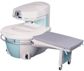
From Philips Medical Systems;
the Panorama 0.23 T, providing a new design optimized for patient comfort, faster reconstruction time than before (300 images/second) and new gradient
specifi cations. Philips' Panorama 0.23 T I/T supports MR-guided interventions, resulting in minimally invasive procedures, more targeted surgery, reduced recovery time and shorter hospital stays. Optional OptoGuide functionality enables real-time needle tracking. Philips' Panorama 0.23 TPanorama 0.2 R/T is the first and only open MRI system to enable radiation therapy planning using MR data sets. The Panorama also features the new and consistent Philips User Interface, an essential element of the Vequion clinical IT family of products and services.
Device Information and Specifi cation CLINICAL APPLICATION Whole body SE, FE, IR, FFE, DEFFE, DESE, TSE, DETSE, Single shot SE, DRIVE, Balanced FFE, MRCP, Fluid Attenuated Inversion Recovery, Turbo FLAIR, IR-TSE, T1-STIR TSE, T2-STIR TSE, Diffusion Imaging, 3D SE, 3D FFE, MTC;; Angiography: CE-ANGIO, MRA 2D, 3D TOFOpen x 46 cm x infinite (side-first patient entry) POWER REQUIREMENTS 400/480 V COOLING SYSTEM TYPE Closed loop chilled water ( chiller included) | | | |
• View the DATABASE results for 'Panorama 0.23T™' (2).
| | | | | | Further Reading: | News & More:
|
|
| |
| | | | | |
| |
|
(PC) Phase contrast sequences are the basis of MRA techniques utilizing the change in the phase shifts of the flowing protons in the region of interest to create an image. Spins that are moving along the direction of a magnetic field gradient receive a phase shift proportional to their velocity.
In a phase contrast sequence two data sets with a different amount of flow sensitivity are acquired. This is usually accomplished by applying gradient pairs, which sequentially dephase and then rephase spins during the sequence. Both 2D and 3D acquisition techniques can be applied with phase contrast MRA.
The first data set is acquired with a flow compensated sequence, i. e. without flow sensitivity. The second data set is acquired with a flow sensitive sequence. The amount of flow sensitivity is controlled by the strength of the bipolar gradient pulse pair, which is incorporated into the sequence. Stationary tissue undergoes no effective phase change after the appli cation of the two gradients. Caused by the different spatial localization of flowing blood to stationary tissue, it experiences a different size of the second bipolar gradient compared to the first. The result is a phase shift.
The raw data from the two data sets are subtracted. By comparing the phase of signals from each lo cation in the two sequences the exact amount of motion induced phase change can be determined to have a map where pixel brightness is proportional to spatial velocity.
Phase contrast images represent the signal intensity of the velocity of spins at each point within the field of view. Regions that are stationary remain black while moving regions are represented as grey to white.
The phase shift is proportional to the spin's velocity, and this allows the quantitative assessment of flow velocities.
The difference MRI signal has a maximum value for opposite directions. This velocity is typically referred to as venc, and depends on the pulse amplitude and distance between the gradient pulse pair. For velocities larger than venc the difference signal is decreased constantly until it gets zero. Therefore, in a phase contrast angiography it is important to correctly set the venc of the sequence to the maximum flow velocity which is expected during the measurement. High venc factors of the PC angiogram (more than 40 cm/sec) will selectively image the arteries ( PCA - arteriography), whereas a venc factor of 20 cm/sec will perform the veins and sinuses (PCV or MRV - venography).
See also Flow Quantification, Contrast Enhanced MR Venography, Time of Flight Angiography, Time Resolved Imaging of Contrast Kinetics. | | | | | |
• View the DATABASE results for 'Phase Contrast Sequence' (5).
| | | | | | Further Reading: | Basics:
|
|
| |
| | | | |
| | |
| | | |
|
| |
 | Look
Ups |
| |