 | Info
Sheets |
| | | | | | | | | | | | | | | | | | | | | | | | |
 | Out-
side |
| | | | |
|
| | | | |
Result : Searchterm 'Cine' found in 5 terms [ ] and 52 definitions [ ] and 52 definitions [ ] ]
| | previous 56 - 57 (of 57) Result Pages : [1] [2 3 4 5 6 7 8 9 10 11 12] |  | |  | Searchterm 'Cine' was also found in the following services: | | | | |
| |  |
| |
|
Magnetic resonance imaging ( MRI) of the spine is a noninvasive procedure to evaluate different types of tissue, including the spinal cord, vertebral disks and spaces between the vertebrae through which the nerves travel, as well as distinguish healthy tissue from diseased tissue.
The cervical, thoracic and lumbar spine MRI should be scanned in individual sections.
The scan protocol parameter like e.g. the field of view ( FOV), slice thickness and matrix are usually different for cervical, thoracic and lumbar spine MRI, but the method
is similar. The standard views in the basic spinal MRI scan to create detailed slices (cross sections) are sagittal T1 weighted and T2 weighted images over the whole body part, and transverse (e.g. multi angle oblique) over the region of interest with different pulse sequences according to the result of the sagittal slices. Additional views or different types of pulse sequences like fat suppression, fluid attenuation inversion recovery ( FLAIR) or
diffusion weighted imaging are created dependent on the indication.
Indications:
•
Neurological deficit, evidence of radiculopathy, cauda equina compression
•
Primary tumors or drop metastases
•
Infection/inflammatory disease, multiple sclerosis
•
Postoperative evaluation of lumbar spine: disk vs. scar
•
Localized back pain with no radiculopathy (leg pain)
Contrast enhanced MRI techniques delineate infections vs. malignancies, show a syrinx cavity and support to differentiate the postoperative conditions. After surgery for disk disease, significant fibrosis can occur in the spine. This scarring can mimic residual disk herniation. Magnetic resonance myelography evaluates spinal stenosis and various intervertebral discs can be imaged with multi angle oblique techniques. Cine series can be used to show true range of motion studies of parts of the spine.
Advanced open MRI devices are developed to perform positional scans in the position of pain or symptom (e.g. Upright™ MRI formerly Stand-Up MRI). | | | | | | | | | | | | |  Further Reading: Further Reading: | | Basics:
|
|
News & More:
| |
| |
| | | Searchterm 'Cine' was also found in the following services: | | | | |
| | |
| |
|
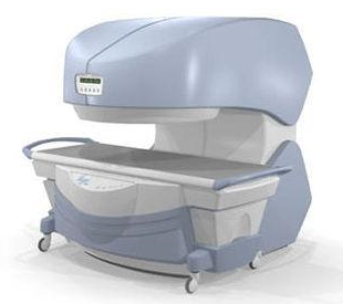
From
Millennium Technology Inc.
This open C-shaped MRI system eases patient comfort and technologist maneuverability. This low cost scanner is build for a wide range of applications. The Virgo™ patient table is detachable and moves on easy rolling castors. Able to accommodate patient weights up to 160 kg, the tabletop has a range of motion of 30 cm in the lateral direction and 90cm in the longitudinal direction. Images generated with this scanner can only be viewed (without data loss) on Millennium's proprietary viewing software.
Device Information and Specification CLINICAL APPLICATION Whole body Head, Body, Neck, Knee, Shoulder,
Spine, Wrist, Breast, Extremity, Lumbar Spine, TMJ
IMAGING MODES Localizer, single slice, multislice, volume, fast, POMP, multi slab, cine, slice and frequency zip, extended dynamic range, tailored RF | | | | | |
| | | | |
| | | |
|
| |
 | Look
Ups |
| |


