 | Info
Sheets |
| | | | | | | | | | | | | | | | | | | | | | | | |
 | Out-
side |
| | | | |
|
| | | | |
Result : Searchterm 'DWI' found in 1 term [ ] and 40 definitions [ ] and 40 definitions [ ] ]
| previous 11 - 15 (of 41) nextResult Pages :  [1] [1]  [2 3 4 5 6 7 8 9] [2 3 4 5 6 7 8 9] |  | |  | Searchterm 'DWI' was also found in the following services: | | | | |
|  |  |
| |
|

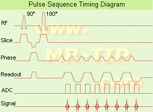
(EPI) Echo planar imaging is one of the early magnetic resonance imaging sequences (also known as Intascan), used in applications like diffusion, perfusion, and functional magnetic resonance imaging. Other sequences acquire one k-space line at each phase encoding step. When the echo planar imaging acquisition strategy is used, the complete image is formed from a single data sample (all k-space lines are measured in one repetition time) of a gradient echo or spin echo sequence (see single shot technique) with an acquisition time of about 20 to 100 ms.
The pulse sequence timing diagram illustrates an echo planar imaging sequence from spin echo type with eight echo train pulses. (See also Pulse Sequence Timing Diagram, for a description of the components.)
In case of a gradient echo based EPI sequence the initial part is very similar to a standard gradient echo sequence. By periodically fast reversing the readout or frequency encoding gradient, a train of echoes is generated.
EPI requires higher performance from the MRI scanner like much larger gradient amplitudes. The scan time is dependent on the spatial resolution required, the strength of the applied gradient fields and the time the machine needs to ramp the gradients.
In EPI, there is water fat shift in the phase encoding direction due to phase accumulations. To minimize water fat shift (WFS) in the phase direction fat suppression and a wide bandwidth (BW) are selected. On a typical EPI sequence, there is virtually no time at all for the flat top of the gradient waveform. The problem is solved by "ramp sampling" through most of the rise and fall time to improve image resolution.
The benefits of the fast imaging time are not without cost. EPI is relatively demanding on the scanner hardware, in particular on gradient strengths, gradient switching times, and receiver bandwidth. In addition, EPI is extremely sensitive to image artifacts and distortions. | |  | | | | | | | | |  Further Reading: Further Reading: | Basics:
|
|
| |
|  | |  |  |  |
| |
|
MRI Contrast Agents:
Contact Information
MAIL
Lantheus Medical Imaging
Bldg. 200-2, 331 Treble Cove Rd.
N. Billerica, MA 01862
USA
| |  | |
• View the DATABASE results for 'Lantheus Medical Imaging, Inc.' (3).
| | |
• View the NEWS results for 'Lantheus Medical Imaging, Inc.' (5).
| | | | |
|  | |  |  |  |
| |
|
A coil is a large inductor with a considerable dimension and a defined wavelength, commonly used in configurations for MR imaging. The frequency of the radio frequency coil is defined by the Larmor relationship. The MRI image quality depends on the signal to noise ratio (SNR) of the acquired signal from the patient. Several MR imaging coils are necessary to handle the diversity of applications. Large coils have a large measurement field, but low signal intensity and vice versa (see also coil diameter). The closer the coil to the object, the stronger the signal - the smaller the volume, the higher the SNR. SNR is very important in obtaining clear images of the human body. The shape of the coil depends on the image sampling. The best available homogeneity can be reached by choice of the appropriate coil type and correct coil positioning. Orientation is critical to the sensitivity of the RF coil and therefore the coil should be perpendicular to the static magnetic field.
RF coils can be differentiated by there function into three general categories:
The RF signal is in the range of 10 to 100 MHz. During a typical set of clinical image measurements, the entire frequency spectrum of interest is of the order 10 kHz, which is an extremely narrow band, considering that the center frequency is about 100 MHz. This allows the use of single-frequency matching techniques for coils because their inherent bandwidth always exceeds the image bandwidth. The multi turn solenoid, bird cage coil, single turn solenoid, and saddle coil are typically operated as the transmitter and receiver of RF energy. The surface and phased array coils are typically operated as a receive only coil.
See also the related poll result: ' 3rd party coils are better than the original manufacturer coils' | | | |  | |
• View the DATABASE results for 'Radio Frequency Coil' (9).
| | | | |  Further Reading: Further Reading: | | Basics:
|
|
News & More:
| |
| |
|  |  | Searchterm 'DWI' was also found in the following services: | | | | |
|  |  |
| |
|

The range of diagnostics and imaging systems of Siemens Medical Systems covers ultrasound, nuclear medicine, angiography, magnetic resonance, computer tomography and patient monitoring. Siemens is one of the three leading MRI manufacturers, which together account for approximately 80 percent of the MRI machines installed worl dwide. Siemens currently offers the Allegra 3T MRI, which is for head scanning only, but the company will also be launching the Trio MRI, a 3T whole body scanner.
Siemens has formed partnerships with more than ten research institutions and private practitioners to define a comprehensive MRI examination and compare MR to currently established cardiovascular modalities, thereby defining optimal diagnosis and treatment.
MRI Scanners:
0.2T to 1.0T:
1.5T:
3.0T to 7.0T:
Hybrid Scanners:
Mobile Solutions:
•
MAGNETOM Espree 1.5T, MAGNETOM Avanto 1.5T and MAGNETOM ESSENZA 1.5T are also offered by Siemens on certified trailers.
Contact Information MAIL
Siemens Medical Solutions
Health Services Corporation
51 Valley Stream Parkway
Malvern, PA 19355
USA | |  | |
• View the DATABASE results for 'Siemens Medical Systems' (14).
| | |
• View the NEWS results for 'Siemens Medical Systems' (3).
| | | | |  Further Reading: Further Reading: | | Basics:
|
|
News & More:
| |
| |
|  | |  |  |  |
| |
|

Image Guidance
Artifacts may appear as a series of fine lines. A narrow bandwidth causes a wide read window, which allows the stimulated echo to be incorporated into the image data. This can be supported by increasing the received bandwidth, which would narrow the read window, thus not incorporating the extraneous echo. Another help would be to change the first echo time, which may change the spacing of the stimulated echoes to outside that of the read window for the second echo. | |  | |
• View the DATABASE results for 'Stimulated Echo' (8).
| | | | |  Further Reading: Further Reading: | Basics:
|
|
| |
|  | |  |  |
|  | | |
|
| |
 | Look
Ups |
| |