Magnetic Resonance - Technology Information Portal Welcome to MRI Technology
Info
  Sheets

Out-
      side


 'Density Weighted Imaging' 
SEARCH FOR    
 
  2 3 5 A B C D E F G H I J K L M N O P Q R S T U V W X Y Z
Result : Searchterm 'Density Weighted Imaging' found in 1 term [] and 1 definition [], (+ 12 Boolean[] results
1 - 5 (of 14)     next
Result Pages : [1]  [2 3]
Searchterm 'Density Weighted Imaging' was also found in the following service: 
spacer
News  (1)  
 
Density Weighted ImagingInfoSheet: - Sequences - 
Intro, 
Overview, 
Types of, 
etc.
 
(PDWI) In density weighted imaging, the contrast is dependent on the density of protons in the tissue. Proton density weighted images are generated by choosing TR greater than T1 (typically ≥ 2 000 ms) and TE less than T2 (typically ≤ 30 ms), the two exponential terms are both close to one and therefore M is relatively independent of T1 and T2, thereby emphasizing Mxy0, which is proportional to the proton density. Also called (Rho) ρ-weighted.

See also Proton Density Weighted Image.
 
Images, Movies, Sliders:
 Knee MRI Coronal Pd Spir 001  Open this link in a new window
    
 MRI - Anatomic Imaging of the Ankle 3  Open this link in a new window
    
SlidersSliders Overview

 
spacer
 
• Share the entry 'Density Weighted Imaging':  Facebook  Twitter  LinkedIn  
 
Further Reading:
  Basics:
IMAGE CONTRAST IN MRI(.pdf)
   by www.assaftal.com    
MRI Resources 
Intraoperative MRI - General - Contrast Agents - Directories - - MR Myelography
 
Proton Density Weighted Image
 
An image produced by controlling the selection of scan parameters to minimize the effects of T1 and T2, resulting in an image dependent primarily on the density of protons in the imaging volume. Proton density contrast is a quantitative summary of the number of protons per unit tissue. The higher the number of protons in a given unit of tissue, the greater the transverse component of magnetization, and the brighter the signal on the proton density contrast image. Conversely the lower the number of protons in a given unit of tissue, the less the transverse magnetization and the darker the signal on the proton density image. Also called (Rho) ρ-weighted.

See also Density Weighted Imaging and Image Contrast Characteristics.
 
Images, Movies, Sliders:
 Knee MRI Coronal Pd Spir 001  Open this link in a new window
    
 
spacer

• View the DATABASE results for 'Proton Density Weighted Image' (4).Open this link in a new window

 
Further Reading:
  Basics:
Musculoskeletal MRI at 3.0 T: Relaxation Times and Image Contrast
Sunday, 1 August 2004   by www.ajronline.org    
Dogs Are People, Too
Saturday, 5 October 2013   by www.nytimes.com    
MRI Resources 
Implant and Prosthesis - MRI Technician and Technologist Jobs - Implant and Prosthesis pool - NMR - Shoulder MRI - MRI Physics
 
Fast Spin EchoForum -
related threadsInfoSheet: - Sequences - 
Intro, 
Overview, 
Types of, 
etc.
 
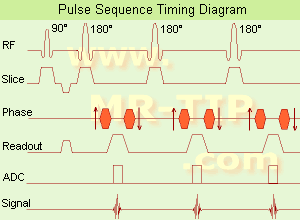
Fast Spin Echo Diagram (FSE) In the pulse sequence timing diagram, a fast spin echo sequence with an echo train length of 3 is illustrated. This sequence is characterized by a series of rapidly applied 180° rephasing pulses and multiple echoes, changing the phase encoding gradient for each echo.
The echo time TE may vary from echo to echo in the echo train. The echoes in the center of the K-space (in the case of linear k-space acquisition) mainly produce the type of image contrast, whereas the periphery of K-space determines the spatial resolution. For example, in the middle of K-space the late echoes of T2 weighted images are encoded. T1 or PD contrast is produced from the early echoes.
The benefit of this technique is that the scan duration with, e.g. a turbo spin echo turbo factor / echo train length of 9, is one ninth of the time. In T1 weighted and proton density weighted sequences, there is a limit to how large the ETL can be (e.g. a usual ETL for T1 weighted images is between 3 and 7). The use of large echo train lengths with short TE results in blurring and loss of contrast. For this reason, T2 weighted imaging profits most from this technique.
In T2 weighted FSE images, both water and fat are hyperintense. This is because the succession of 180° RF pulses reduces the spin spin interactions in fat and increases its T2 decay time. Fast spin echo (FSE) sequences have replaced conventional T2 weighted spin echo sequences for most clinical applications. Fast spin echo allows reduced acquisition times and enables T2 weighted breath hold imaging, e.g. for applications in the upper abdomen.
In case of the acquisition of 2 echoes this type of a sequence is named double fast spin echo / dual echo sequence, the first echo is usually density and the second echo is T2 weighted image. Fast spin echo images are more T2 weighted, which makes it difficult to obtain true proton density weighted images. For dual echo imaging with density weighting, the TR should be kept between 2000 - 2400 msec with a short ETL (e.g., 4).
Other terms for this technique are:
Turbo Spin Echo
Rapid Imaging Spin Echo,
Rapid Spin Echo,
Rapid Acquisition Spin Echo,
Rapid Acquisition with Refocused Echoes
 
Images, Movies, Sliders:
 Lumbar Spine T2 FSE Sagittal  Open this link in a new window
    

Courtesy of  Robert R. Edelman
 MRI - Anatomic Imaging of the Foot  Open this link in a new window
    
SlidersSliders Overview

 Lumbar Spine T2 FSE Axial  Open this link in a new window
    

Courtesy of  Robert R. Edelman
 
spacer

• View the DATABASE results for 'Fast Spin Echo' (31).Open this link in a new window

 
Further Reading:
  Basics:
MYELIN-SELECTIVE MRI: PULSE SEQUENCE DESIGN AND OPTIMIZATION
   by www.imaging.robarts.ca    
Advances in Magnetic Resonance Neuroimaging
Friday, 27 February 2009   by www.ncbi.nlm.nih.gov    
  News & More:
New MR sequence helps radiologists more accurately evaluate abnormalities of the uterus and ovaries
Thursday, 23 April 2009   by www.eurekalert.org    
Spin echoes, CPMG and T2 relaxation - Introductory NMR & MRI from Magritek
2013   by www.azom.com    
Searchterm 'Density Weighted Imaging' was also found in the following service: 
spacer
News  (1)  
 
Fast Imaging with Steady State PrecessionInfoSheet: - Sequences - 
Intro, 
Overview, 
Types of, 
etc.
 
(FISP) A fast imaging sequence, which attempts to combine the signals observed separately in the FADE sequence, generally sensitive about magnetic susceptibility artifacts and imperfections in the gradient waveforms. Confusingly now often used to refer to a refocused FLASH type sequence.
This sequence is very similar to FLASH, except that the spoiler pulse is eliminated. As a result, any transverse magnetization still present at the time of the next RF pulse is incorporated into the steady state. FISP uses a RF pulse that alternates in sign. Because there is still some remaining transverse magnetization at the time of the RF pulse, a RF pulse of a degree flips the spins less than a degree from the longitudinal axis. With small flip angles, very little longitudinal magnetization is lost and the image contrast becomes almost independent of T1. Using a very short TE (with TR 20-50 ms, flip angle 30-45°) eliminates T2* effects, so that the images become proton density weighted. As the flip angle is increased, the contrast becomes increasingly dependent on T1 and T2*. It is in the domain of large flip angles and short TR that FISP exhibits vastly different contrast to FLASH type sequences. Used for T1 orthopedic imaging, 3D MPR, cardiography and angiography.
spacer

• View the DATABASE results for 'Fast Imaging with Steady State Precession' (5).Open this link in a new window

 
Further Reading:
  Basics:
MRI techniques improve pulmonary embolism detection
Monday, 19 March 2012   by medicalxpress.com    
MRI Resources 
Intraoperative MRI - Research Labs - Safety pool - Movies - Stimulator pool - MRA
 
Fast Low Angle ShotInfoSheet: - Sequences - 
Intro, 
Overview, 
Types of, 
etc.MRI Resource Directory:
 - Sequences -
 
(FLASH) A fast sequence producing signals called gradient echo with low flip angles. FLASH sequences are modifications, which incorporate or remove the effects of transverse coherence respectively.
FLASH uses a semi-random spoiler gradient after each echo to spoil the steady state (to destroy any remaining transverse magnetization) by causing a spatially dependent phase shift. The transverse steady state is spoiled but the longitudinal steady state depends on the T1 values and the flip angle. Extremely short TR times are possible, as a result the sequence provides a mechanism for gaining extremely high T1 contrast by imaging with TR times as brief as 20 to 30 msec while retaining reasonable signal levels. It is important to keep the TE as short as possible to suppress susceptibility artifacts.
The T1 contrast depends on the TR as well as on flip angle, with short TE.
Small flip angles and short TR results in proton density, and long TR in T2* weighting.
With large flip angles and short TR result T1 weighted images.

TR and flip angle adjustment:

TR 3000 ms, Flip Angle 90°
TR 1500 ms, Flip Angle 45°
TR 700 ms, Flip Angle 25°
TR 125 ms, Flip Angle 10°

The apparent ability to trade TR against flip angle for purposes of contrast and the variation in SNR as the scan time (TR) is reduced.

See also Gradient Echo Sequence.
 
Images, Movies, Sliders:
 Fetus (Brain) and Dermoid in Mother  Open this link in a new window
      

Courtesy of  Robert R. Edelman

 
spacer

• View the DATABASE results for 'Fast Low Angle Shot' (5).Open this link in a new window

 
Further Reading:
  News & More:
Motion Compensation in MR Imaging
   by ccn.ucla.edu    
Turbo-FLASH Based Arterial Spin Labeled Perfusion MRI at 7 T
Thursday, 20 June 2013   by www.plosone.org    
Usefulness of MR Imaging for Diseases of the Small Intestine: Comparison with CT
2000   by www.ncbi.nlm.nih.gov    
MRI Resources 
Abdominal Imaging - Nerve Stimulator - Spine MRI - Intraoperative MRI - Image Quality - Hospitals
 
     1 - 5 (of 14)     next
Result Pages : [1]  [2 3]
 Random Page
 
Share This Page
FacebookTwitterLinkedIn

MR-TIP    
Community   
User
Pass
Forgot your UserID/Password ?    


How AI will impact MRI :
only diagnostics 
saving time 
reducing cost 
makes planning obsolete 
reduce human knowledge 
not at all 

Look
      Ups


MR-TIP.com uses cookies! By browsing MR-TIP.com, you agree to our use of cookies.

Magnetic Resonance - Technology Information Portal
Member of SoftWays' Medical Imaging Group - MR-TIP • Radiology-TIP • Medical-Ultrasound-Imaging • 
Copyright © 2003 - 2024 SoftWays. All rights reserved. [ 21 November 2024]
Terms of Use | Privacy Policy | Advertising
 [last update: 2024-02-26 03:41:00]