 | Info
Sheets |
| | | | | | | | | | | | | | | | | | | | | | | | |
 | Out-
side |
| | | | |
|
| | | 'Diffusion Tensor Imaging' | |
Result : Searchterm 'Diffusion Tensor Imaging' found in 1 term [ ] and 8 definitions [ ] and 8 definitions [ ], (+ 1 Boolean[ ], (+ 1 Boolean[ ] results ] results
| previous 6 - 10 (of 10) Result Pages :  [1] [1]  [2] [2] |  | |  | Searchterm 'Diffusion Tensor Imaging' was also found in the following services: | | | | |
|  |  |
| |
|

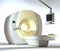
From Philips Medical Systems;
the Intera-family offers with this member a wide range of possibilities, efficiency and a ergonomic and intuitive serving-platform. Also available as Intera CV for cardiac and Intera I/T for interventional MR procedures.
The scanners are also equipped with SENSE technology, which is essential for high-quality contrast enhanced magnetic resonance angiography, interactive cardiac MR and diffusion tensor imaging ( DTI) fiber tracking.
The increased accuracy and clarity of MR scans obtained with this technology allow for faster and more accurate diagnosis of potential problems like patient friendliness and expands the breadth of applications including cardiology, oncology and interventional MR.
Device Information and Specification
CLINICAL APPLICATION
Whole body
CONFIGURATION
Short bore compact
Standard: head, body, C1, C3; Optional: Small joint, flex-E, flex-R, endocavitary (L and S), dual TMJ, knee, neck, T/L spine, breast; Optional phased array: Spine, pediatric, 3rd party connector; Optional SENSE coils: Flex-S-M-L, flex body, flex cardiac
SE, Modified-SE ( TSE), IR (T1, T2, PD), STIR, FLAIR, SPIR, FFE, T1-FFE, T2-FFE, Balanced FFE, TFE, Balanced TFE, Dynamic, Keyhole, 3D, Multi Chunk 3D, Multi Stack 3D, K Space Shutter, MTC, TSE, Dual IR, DRIVE, EPI, Cine, 2DMSS, DAVE, Mixed Mode; Angiography: PCA, MCA, Inflow MRA, CE
TR
2.9 (Omni), 1.6 (Power), 1.6 (Master/Expl) msec
TE
1.0 (Omni), 0.7 (Power), 0.5 (Master/Expl) msec
RapidView Recon. greater than 500 @ 256 Matrix
0.1 mm(Omni), 0.05 mm (Pwr/Mstr/Expl)
128 x 128, 256 x 256,512 x 512,1024 x 1024 (64 for BOLD img.)
Variable in 1% increments
Lum.: 120 cd/m2; contrast: 150:1
Variable (op. param. depend.)
POWER REQUIREMENTS
380/400 V
| |  | | | | | | | | |
|  | |  |  |  |
| |
|

From Philips Medical Systems;
the Intera 3 T high field system, the first with a compact magnet, which is built on the same platform as the 1.5 T, is targeted to high-end neurological, orthopedic and cardiovascular imaging applications with maximum patient comfort and acceptance without compromising image quality and clinical performance. Useable for clinical routine and research.
The Intera systems offer diffusion tensor imaging ( DTI) fiber tracking that measures movement of water in the brain and can therefore detect areas of the brain where normal movement of water is disrupted.
Device Information and Specification
CLINICAL APPLICATION
Whole body
CONFIGURATION
Short bore compact
Standard: head, body, C1, C3; Optional: Small joint, flex-E, flex-R, endocavitary (L and S), dual TMJ, knee, neck, T/L spine, breast; Optional phased array: spine;; Optional SENSE coils: Flex body, flex cardiac, neuro-vascular, head
SE, Modified-SE, IR (T1, T2, PD), STIR, FLAIR, SPIR, FFE, T1-FFE, T2-FFE, Balanced FFE, TFE, Balanced TFE, Dynamic, Keyhole, 3D, Multi Chunk 3D, Multi Stack 3D, K Space Shutter, MTC, TSE, Dual IR, DRIVE, EPI, Cine, 2DMSS, DAVE, Mixed Mode; Angiography: Inflow MRA, TONE, PCA, CE MRA
TR
Min. 1.6 (Master) msec
TE
Min. 0.5 (Master) msec
RapidView Recon. greater than 500 @ 256 Matrix
0.1 mm (Omni), 0.05 mm (Power)
128 x 128, 256 x 256,512 x 512,1024 x 1024 (64 for Bold img)
Variable in 1% increments
Lum.: 120 cd/m2; contrast: 150:1
Variable (op. param. depend.)
POWER REQUIREMENTS
380/400 V
STRENGTH
30 (Master) mT/m
| |  | |
• View the DATABASE results for 'Intera 3.0T™' (2).
| | | | |
|  | |  |  |  |
| |
|
Rapid echo planar imaging and high-performance MRI gradient systems create fast-switching magnetic fields that can stimulate muscle and nerve tissues produced by either changing the electrical resistance or the potential of the excitation. There are apparently no effects on the conduction of impulses in the nerve fiber up to field strength of 0.1 T. A preliminary study has indicated neurological effects by exposition to a whole body imager at 4.0 T. Theoretical examinations argue that field strengths of 24 T are required to produce a 10% reduction of nerve impulse conduction velocity.
Nerve stimulations during MRI scans can be induced by very rapid changes of the magnetic field. This stimulation may occur for example during diffusion weighted sequences or diffusion tensor imaging and can result in muscle contractions caused by effecting motor nerves. The so-called magnetic phosphenes are attributed to magnetic field variations and may occur in a threshold field change of between 2 and 5 T/s. Phosphenes are stimulations of the optic nerve or the retina, producing a flashing light sensation in the eyes. They seem not to cause any damage in the eye or the nerve.
Varying magnetic fields are also used to stimulate bone-healing in non-unions and pseudarthroses. The reasons why pulsed magnetic fields support bone-healing are not completely understood. The mean threshold levels for various stimulations are 3600 T/s for the heart, 900 T/s for the respiratory system, and 60 T/s for the peripheral nerves.
Guidelines in the United States limit switching rates at a factor of three below the mean threshold for peripheral nerve stimulation. In the event that changes in nerve conductivity happens, the MRI scan parameters should be adjusted to reduce dB/dt for nerve stimulation. | |  | |
• View the DATABASE results for 'Nerve Conductivity' (2).
| | | | |  Further Reading: Further Reading: | | Basics:
|
|
News & More:
| |
| |
|  |  | Searchterm 'Diffusion Tensor Imaging' was also found in the following services: | | | | |
|  |  |
| |
|
Special imaging primarily means advanced MRI techniques used for qualitative and quantitative measurement of biological metabolism as e.g., spectroscopy, perfusion imaging (PWI, ASL), diffusion weighted imaging ( DWI, DTI, DTT) and brain function ( BOLD, fMRI). This physiological magnetic resonance techniques offer insights into brain structure, function, and metabolism.
Spectroscopy provides functional information related to identification and quantification of e.g. brain metabolites.
MR perfusion imaging has applications in stroke, trauma, and brain neoplasm. MRI provides the high spatial and temporal resolution needed to measure blood flow to the brain. arterial spin labeling techniques utilize the intrinsic protons of blood and brain tissue, labeled by special preparation pulses, rather than exogenous tracers injected into the blood.
MR diffusion tensor imaging characterizes the ability of water to spread across the brain in different directions. Diffusion parallel to nerve fibers has been shown to be greater than diffusion in the perpendicular direction. This provides a tool to study in vivo fiber connectivity in brain MRI.
FMRI allows the detection of a functional activation in the brain because cortical activity is intimately related to local metabolism changes. See also Diffusion Tensor Tractography. | |  | |
• View the NEWS results for 'Special Imaging' (14).
| | | | |  Further Reading: Further Reading: | | Basics:
|
|
News & More:
| |
| |
|  | |  |  |  |
| |
|
(ADC) A diffusion coefficient to differentiate T2 shine through effects or artifacts from real ischemic lesions. In the human brain, water diffusion is a three-dimensional process that is not truly random because the diffusional motion of water is impeded by natural barriers. These barriers are cell membranes, myelin sheaths, white matter fiber tracts, and protein molecules.
The apparent water diffusion coefficients can be calculated by acquiring two or more images with a different gradient duration and amplitude (b-values). The contrast in the ADC map depends on the spatially distributed diffusion coefficient of the acquired tissues and does not contain T1 and T2* values.
The increased sensitivity of diffusion-weighted MRI in detecting acute ischemia is thought to be the result of the water shift intracellularly restricting motion of water protons (cytotoxic edema), whereas the conventional T2 weighted images show signal alteration mostly as a result of vasogenic edema.
The reduced ADC value also could be the result of decreased temperature in the nonperfused tissues, loss of brain pulsations leading to a decrease in apparent proton motion, increased tissue osmolality associated with ischemia, or a combination of these factors.
The lower ADC measurements seen with early ischemia, have not been fully established, however, a lower apparent ADC is a sensitive indicator of early ischemic brain at a stage when ischemic tissue remains potentially salvageable.
See also Diffusion Weighted Imaging and Diffusion Tensor Tractography. | |  | |
• View the DATABASE results for 'Apparent Diffusion Coefficient' (4).
| | | | |  Further Reading: Further Reading: | Basics:
|
|
News & More:
| |
| |
|  | |  |  |
|  | previous 6 - 10 (of 10) Result Pages :  [1] [1]  [2] [2] |
| |
|
| |
 | Look
Ups |
| |