 | Info
Sheets |
| | | | | | | | | | | | | | | | | | | | | | | | |
 | Out-
side |
| | | | |
|
| | | 'Diffusion Weighted Whole Body Imaging' | |
Result : Searchterm 'Diffusion Weighted Whole Body Imaging' found in 1 term [ ] and 0 definition [ ] and 0 definition [ ], (+ 5 Boolean[ ], (+ 5 Boolean[ ] results ] results
| 1 - 5 (of 6) nextResult Pages :  [1] [1]  [2] [2] |  | | |  |  |  |
| Diffusion Weighted Whole Body Imaging | |
| |
|
(DWIBS) This diffusion weighted whole body imaging with background signal suppression provides diffusion weighted contrast throughout the body using a single or multi-station background-suppressed diffusion imaging. DWIBS images are comparable with PET images and provide applications from the visualization of nerve roots and brachial plexus to the detection of lesions throughout the body. See also Diffusion Weighted Imaging (DWI). | |  | | | | • Share the entry 'Diffusion Weighted Whole Body Imaging':    | | | | |  Further Reading: Further Reading: | | Basics:
|
|
News & More:
| |
| |
|  | |  |  |  |
| |
|
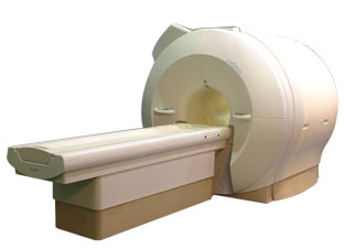
Device Information and Specification
CLINICAL APPLICATION
Whole body
CONFIGURATION
Mobile compact
Whole body, intra-operative head, neck volume, atlas head//neck vascular quadrature phased array, spine quadrature, C/T/L spine phased array, small joint, large joint, TMJ bilateral, shoulder phased array, extremity quadrature volume, wrist, hand quadrature, general purpose flexible, pelvis/abdomen phased array, body quadrature, phased array flexible, breast bilateral
IMAGING MODES
Localizer, single slice, multislice, volume
| |  | |
• View the DATABASE results for 'iMotion™ 1.5 Tesla Magnet' (2).
| | | | |
|  | |  |  |  |
| |
|

'Next generation MRI system 1.5T CHORUS developed by ISOL Technology is optimized for both clinical diagnostic imaging and for research development.
CHORUS offers the complete range of feature oriented advanced imaging techniques- for both clinical routine and research. The compact short bore magnet, the patient friendly design and the gradient technology make the innovation to new degree of perfection in magnetic resonance.'
Device Information and Specification
CLINICAL APPLICATION
Whole body
Spin Echo, Gradient Echo, Fast Spin Echo,
Inversion Recovery ( STIR, Fluid Attenuated Inversion Recovery), FLASH, FISP, PSIF, Turbo Flash ( MPRAGE ),TOF MR Angiography, Standard echo planar imaging package (SE-EPI, GE-EPI), Optional:
Advanced P.A. Imaging Package (up to 4 ch.), Advanced echo planar imaging package,
Single Shot and Diffusion Weighted EPI, IR/FLAIR EPI
STRENGTH
20 mT/m (Upto 27 mT/m)
| |  | |
• View the DATABASE results for 'CHORUS 1.5T™' (2).
| | | | |
|  | |  |  |  |
| |
|
(DWI) Magnetic resonance imaging is sensitive to diffusion, because the diffusion of water molecules along a field gradient reduces the MR signal. In areas of lower diffusion the signal loss is less intense and the display from this areas is brighter. The use of a bipolar gradient pulse and suitable pulse sequences permits the acquisition of diffusion weighted images (images in which areas of rapid proton diffusion can be distinguished from areas with slow diffusion).
Based on echo planar imaging, multislice DWI is today a standard for imaging brain infarction. With enhanced gradients, the whole brain can be scanned within seconds. The degree of diffusion weighting correlates with the strength of the diffusion gradients, characterized by the b-value, which is a function of the gradient related parameters: strength, duration, and the period between diffusion gradients.
Certain illnesses show restrictions of diffusion, for example demyelinization and cytotoxic edema. Areas of cerebral infarction have decreased apparent diffusion, which results in increased signal intensity on diffusion weighted MRI scans. DWI has been demonstrated to be more sensitive for the early detection of stroke than standard pulse sequences and is closely related to temperature mapping.
DWIBS is a new diffusion weighted imaging technique for the whole body that produces PET-like images. The DWIBS sequence has been developed with the aim to detect lymph nodes and to differentiate normal and hyperplastic from metastatic lymph nodes. This may be possible caused by alterations in microcirculation and water diffusivity within cancer metastases in lymph nodes.
See also Diffusion Weighted Sequence, Perfusion Imaging, ADC Map, Apparent Diffusion Coefficient, and Diffusion Tensor Imaging. | |  | |
• View the DATABASE results for 'Diffusion Weighted Imaging' (11).
| | |
• View the NEWS results for 'Diffusion Weighted Imaging' (4).
| | | | |  Further Reading: Further Reading: | Basics:
|
|
News & More:
| |
| |
|  | |  |  |  |
| |
|
Magnetic resonance imaging ( MRI) of the spine is a noninvasive procedure to evaluate different types of tissue, including the spinal cord, vertebral disks and spaces between the vertebrae through which the nerves travel, as well as distinguish healthy tissue from diseased tissue.
The cervical, thoracic and lumbar spine MRI should be scanned in individual sections.
The scan protocol parameter like e.g. the field of view ( FOV), slice thickness and matrix are usually different for cervical, thoracic and lumbar spine MRI, but the method
is similar. The standard views in the basic spinal MRI scan to create detailed slices (cross sections) are sagittal T1 weighted and T2 weighted images over the whole body part, and transverse (e.g. multi angle oblique) over the region of interest with different pulse sequences according to the result of the sagittal slices. Additional views or different types of pulse sequences like fat suppression, fluid attenuation inversion recovery ( FLAIR) or
diffusion weighted imaging are created dependent on the indication.
Indications:
•
Neurological deficit, evidence of radiculopathy, cauda equina compression
•
Primary tumors or drop metastases
•
Infection/inflammatory disease, multiple sclerosis
•
Postoperative evaluation of lumbar spine: disk vs. scar
•
Localized back pain with no radiculopathy (leg pain)
Contrast enhanced MRI techniques delineate infections vs. malignancies, show a syrinx cavity and support to differentiate the postoperative conditions. After surgery for disk disease, significant fibrosis can occur in the spine. This scarring can mimic residual disk herniation. Magnetic resonance myelography evaluates spinal stenosis and various intervertebral discs can be imaged with multi angle oblique techniques. Cine series can be used to show true range of motion studies of parts of the spine.
Advanced open MRI devices are developed to perform positional scans in the position of pain or symptom (e.g. Upright™ MRI formerly Stand-Up MRI). | | | |  | |
• View the DATABASE results for 'Spine MRI' (11).
| | |
• View the NEWS results for 'Spine MRI' (4).
| | | | |  Further Reading: Further Reading: | Basics:
|
|
News & More:
| |
| |
|  | |  |  |
|  | 1 - 5 (of 6) nextResult Pages :  [1] [1]  [2] [2] |
| |
|
| |
 | Look
Ups |
| |