 | Info
Sheets |
| | | | | | | | | | | | | | | | | | | | | | | | |
 | Out-
side |
| | | | |
|
| | | | | |  | Searchterm 'Excitation' was also found in the following services: | | | | |
|  |  |
| |
|
Quick Overview
Please note that there are different common names for this MRI artifact.
DESCRIPTION
Image wrap around
Aliasing is an artifact that occurs in MR images when the scanned body part is larger than field of view ( FOV). As a consequence of the acquired k-space frequencies not being sampled densely enough, whereby portions of the object outside of the desired FOV get mapped to an incorrect location inside the FOV. The cyclical property of the Fourier transform fills the missing data of the right side with data from behind the FOV of the left side and vice versa. This is caused by a too small number of samples acquired in, e.g. the frequency encoding direction, therefore the spectrums will overlap, resulting in a replication of the object in the x direction.
Aliasing in the frequency direction can be eliminated by twice as fast sampling of the signal or by applying frequency specific filters to the received signal.
A similar problem occurs in the phase encoding direction, where the phases of signal-bearing tissues outside of the FOV in the y-direction are a replication of the phases that are encoded within the FOV. Phase encoding gradients are scaled for the field of view only, therefore tissues outside the FOV do not get properly phase encoded relative to their actual position and 'wraps' into the opposite side of the image.

Image Guidance
| |  | | | | | | | | |
|  |  | Searchterm 'Excitation' was also found in the following services: | | | | |
|  |  |
| |
|

From Aurora Imaging Technology, Inc.;
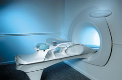
The Aurora® 1.5T Dedicated Breast MRI System with Bilateral SpiralRODEO™ is the first and only FDA approved MRI device designed specifically for breast imaging. The Aurora System, which is already in clinical use at a growing number of leading breast care centers in the US, Europe, got in December 2006 also the approval from the State Food and Drug Administration of the People's Republic of China (SFDA).
'Some of the proprietary and distinguishing features of the Aurora System include: 1) an ellipsoid magnetic shim that provides coverage of both breasts, the chest wall and bilateral axillary lymph nodes; 2) a precision gradient coil with the high linearity required for high resolution spiral reconstruction;; 3) a patient-handling table that provides patient comfort and procedural utility; 4) a fully integrated Interventional System for MRI guided biopsy and localization; and 5) the user-friendly AuroraCAD™ computer-aided image display system designed to improve the accuracy and efficiency of diagnostic interpretations.'
Device Information and Specification
CONFIGURATION
Short bore compact
TE
From 5 ms for RODEO Plus to over 80 ms, 120 ms for T2 sequences
Around 0.02 sec for a 256x256 image, 12.4 sec for a 512 x 512 x 32 multislice set
20 - 36 cm, max. elliptical 36 x 44 cm
POWER REQUIREMENTS
150A/120V-208Y/3 Phase//60 Hz/5 Wire
| |  | |
• View the DATABASE results for 'Aurora® 1.5T Dedicated Breast MRI System' (2).
| | |
• View the NEWS results for 'Aurora® 1.5T Dedicated Breast MRI System' (3).
| | | | |  Further Reading: Further Reading: | News & More:
|
|
| |
|  | |  |  |  |
| |
|
(BW) Bandwidth is a measure of frequency range, the range between the highest and lowest frequency allowed in the signal. For analog signals, which can be mathematically viewed as a function of time, bandwidth is the width, measured in Hertz of a frequency range in which the signal's Fourier transform is nonzero.

Image Guidance
| |  | |
• View the DATABASE results for 'Bandwidth' (19).
| | | | |  Further Reading: Further Reading: | | Basics:
|
|
News & More:
| |
| |
|  |  | Searchterm 'Excitation' was also found in the following services: | | | | |
|  |  |
| |
|
Brain imaging, magnetic resonance imaging of the head or skull, cranial magnetic resonance tomography (MRT), neurological MRI - they describe all the same radiological imaging technique for medical diagnostic.
Magnetic resonance imaging of the human brain includes the anatomic description and the detection of lesions. Special techniques like diffusion weighted imaging, functional magnetic resonance imaging ( fMRI) and spectroscopy provide also information about the function and chemical metabolites of the brain.
MRI provides detailed pictures of brain and nerve tissues in multiple planes without obstruction by overlying bones. Brain MRI is the procedure of choice for most brain disorders. It provides clear images of the brainstem and posterior brain, which are difficult to view on a CT scan. It is also useful for the diagnosis of demyelinating disorders (disorders such as multiple sclerosis (MS) that cause destruction of the myelin sheath of the nerve).
With this noninvasive procedure also the evaluation of blood flow and the flow of cerebrospinal fluid (CSF) is possible. Different MRA methods, also without contrast agents can show a venous or arterial angiogram. MRI can distinguish tumors, inflammatory lesions, and other pathologies from the normal brain anatomy. However, MRI scans are also used instead other methods to avoid the dangers of interventional procedures like angiography (DSA - digital subtraction angiography) as well as of repeated exposure to radiation as required for computed tomography (CT) and other X-ray examinations.
A ( birdcage) bird cage coil achieves uniform excitation and reception and is commonly used to study the brain. Usually a brain MRI procedure includes FLAIR, T2 weighted and T1 weighted sequences in two or three planes. See also Fetal MRI, Fluid Attenuation Inversion Recovery ( FLAIR), Perfusion Imaging and High Field MRI. See also Arterial Spin Labeling. | | | | | | | |
• View the DATABASE results for 'Brain MRI' (14).
| | |
• View the NEWS results for 'Brain MRI' (32).
| | | | |  Further Reading: Further Reading: | | Basics:
|
|
News & More:
|  |
MRI Reveals Significant Brain Abnormalities Post-COVID
Monday, 21 November 2022 by neurosciencenews.com |  |  |
Combining genetics and brain MRI can aid in predicting chances of Alzheimer's disease
Wednesday, 29 June 2022 by www.sciencedaily.com |  |  |
Roundup: How Even Mild COVID Can Affect the Brain; This Many Daily Steps Improves Longevity; and More
Friday, 11 March 2022 by baptisthealth.net |  |  |
A low-cost and shielding-free ultra-low-field brain MRI scanner
Tuesday, 14 December 2021 by www.nature.com |  |  |
Large International Study Reveals Spectrum of COVID-19 Brain Complications
Tuesday, 9 November 2021 by www.itnonline.com |  |  |
Brain MRI-Based Subtypes of MS Predict Disability Progression, Treatment Response
Thursday, 13 May 2021 by www.neurologyadvisor.com |  |  |
New MRI method improves detection of disease changes in the brain's network
Thursday, 11 June 2020 by www.compute.dtu.dk |  |  |
New NeuroCOVIDÂť Classification System Uses MRI to Categorize Patients
Friday, 12 June 2020 by www.diagnosticimaging.com |  |  |
New MRI technique can 'see' molecular changes in the brain
Thursday, 5 September 2019 by medicalxpress.com |  |  |
Talking therapy or medication for depression: Brain scan may help suggest better treatment
Monday, 27 March 2017 by www.newsnation.in |  |  |
MRI identifies brain abnormalities in chronic fatigue syndrome patients
Wednesday, 29 October 2014 by www.eurekalert.org |  |  |
MRIs Useful in Tracking Depression in MS Patients
Tuesday, 1 July 2014 by www.hcplive.com |  |  |
Contrast agent linked with brain abnormalities on MRI
Tuesday, 17 December 2013 by www.sciencecodex.com |  |  |
MRIs Reveal Signs of Brain Injuries Not Seen in CT Scans
Tuesday, 18 December 2012 by www.sciencedaily.com |  |  |
Iron Deposits in the Brain May Be Early Indicator of MS
Wednesday, 13 November 2013 by www.healthline.com |  |  |
Migraine Sufferers Have Thicker Brain Cortex
Tuesday, 20 November 2007 by www.medicalnewstoday.com |
|
| |
|  |  | Searchterm 'Excitation' was also found in the following services: | | | | |
|  |  |
| |
|
(CSI) Chemical shift imaging is an extension of MR spectroscopy, allowing metabolite information to be measured in an extended region and to add the chemical analysis of body tissues to the potential clinical utility of Magnetic Resonance. The spatial location is phase encoded and a spectrum is recorded at each phase encoding step to allow the spectra acquisition in a number of volumes covering the whole sample. CSI provides mapping of chemical shifts, analog to individual spectral lines or groups of lines.
Spatial resolution can be in one, two or three dimensions, but with long acquisition times od full 3D CSI. Commonly a slice-selected 2D acquisition is used. The chemical composition of each voxel is represented by spectra, or as an image in which the signal intensity depends on the concentration of an individual metabolite. Alternatively frequency-selective pulses excite only a single spectral component.
There are several methods of performing chemical shift imaging, e.g. the inversion recovery method, chemical shift selective imaging sequence, chemical shift insensitive slice selective RF pulse, the saturation method, spatial and chemical shift encoded excitation and quantitative chemical shift imaging.
See also Magnetic Resonance Spectroscopy. | |  | |
• View the DATABASE results for 'Chemical Shift Imaging' (6).
| | | | |  Further Reading: Further Reading: | Basics:
|
|
News & More:
| |
| |
|  | |  |  |
|  | | |
|
| |
 | Look
Ups |
| |