Magnetic Resonance - Technology Information Portal Welcome to MRI Technology
Info
  Sheets

Out-
      side


 'Flow' 
SEARCH FOR    
 
  2 3 5 A B C D E F G H I J K L M N O P Q R S T U V W X Y Z
Result : Searchterm 'Flow' found in 15 terms [] and 98 definitions []
previous     51 - 55 (of 113)     next
Result Pages : [1 2 3]  [4 5 6 7 8 9 10 11 12 13 14 15 16 17 18 19 20 ... ]
Searchterm 'Flow' was also found in the following services: 
spacer
News  (51)  Resources  (20)  Forum  (14)  
 
2 Dimensional Time of Flight Magnetic Resonance AngiographyInfoSheet: - Sequences - 
Intro, 
Overview, 
Types of, 
etc.MRI Resource Directory:
 - MRA -
 
(2D TOF MRA) This form of MR angiography is based on the acquisition of multiple, short-TR, gradient echo single slice images. 2D TOF MRA is the preferred technique for visualizing slow flow, how for example it happens in veins. 2D TOF MRA consists of multiple sequentially-acquired single slices, therefore the saturation effects are minimized.
spacer
Searchterm 'Flow' was also found in the following services: 
spacer
Radiology  (25) Open this link in a new windowUltrasound  (120) Open this link in a new window
Ampere
 
(A or amp) The SI base unit of electric current.
Definition: Two parallel conductors, infinitely long and having negligible cross section, should be placed 1 meter apart in a perfect vacuum. One ampere is the current that creates between them a force of 0.2 micronewton per meter of length.
One ampere represents a current flow of 1 coulomb of charge per second.
One ampere of current results from a potential distribution of 1 volt per ohm of resistance, or from a power production rate of 1 watt per volt of potential.
The unit is known informally as the amp, but A is its official symbol and is named for the French physicist André-Marie Ampère.
spacer

• View the DATABASE results for 'Ampere' (8).Open this link in a new window

 
Further Reading:
  Basics:
How Many? A Dictionary of Units of Measurement © Russ Rowlett and the University of North Carolina at Chapel Hill
Wednesday, 21 March 2001   by www.unc.edu    
MRI Resources 
Spectroscopy - DICOM - MRI Accidents - MR Guided Interventions - Safety pool - Spine MRI
 
AngiographyForum -
related threads
 
Angiography means the imaging of veins and arteries. Magnetic resonance angiography (MRA) has a lower invasion than conventional angiography with catheter and X-ray contrast agent. Time of flight angiography (inflow) and phase contrast angiography works without contrast agents. Only in contrast enhanced magnetic resonance angiography is the use of contrast agents necessary, but the lack of side effects is an advantage of MRI contrast agents, just as the smaller dosage as used in X-ray angiography techniques.

See also the related poll result: 'MRI will have replaced 50% of x-ray exams by'
 
Images, Movies, Sliders:
 CE-MRA of the Carotid Arteries Colored MIP  Open this link in a new window
    
SlidersSliders Overview

 CE MRA of the Aorta  Open this link in a new window
    
SlidersSliders Overview

 TOF-MRA Circle of Willis Inverted MIP  Open this link in a new window


spacer

• View the DATABASE results for 'Angiography' (120).Open this link in a new window


• View the NEWS results for 'Angiography' (15).Open this link in a new window.
 
Further Reading:
  News & More:
Usefulness of non-contrast-enhanced magnetic resonance imaging prior to venous interventions
Tuesday, 18 September 2018   by www.ncbi.nlm.nih.gov    
Searchterm 'Flow' was also found in the following services: 
spacer
News  (51)  Resources  (20)  Forum  (14)  
 
Black Boundary ArtifactInfoSheet: - Artifacts - 
Case Studies, 
Reduction Index, 
etc.MRI Resource Directory:
 - Artifacts -
 
Quick Overview
Please note that there are different common names for this artifact.
Artifact Information
NAME
Black boundary, dark boundary, contour, chemical shift, relief
DESCRIPTION
Black contours at boundaries
Black boundary artifacts are black lines following voxels where both water and fat protons are present in the same voxel. This artifact arise along the boundary of organs or tissues perpendicular to the frequency encoding direction, and occurs preferentially in gradient echo sequences with out of phase echo times.
mri safety guidance
Image Guidance
Fat suppression techniques eliminate this artifact. For artifact reducing helps a smaller water fat shift (high bandwidth), a higher matrix or/and an in phase TE.

See also Chemical Shift Artifact.
spacer

• View the DATABASE results for 'Black Boundary Artifact' (4).Open this link in a new window

 
Further Reading:
  Basics:
What is chemical shift artefact? Why does it occur? How many Hz at 1.5 T?
   by www.revisemri.com    
Searchterm 'Flow' was also found in the following services: 
spacer
Radiology  (25) Open this link in a new windowUltrasound  (120) Open this link in a new window
CHORUS 1.5Tâ„¢InfoSheet: - Devices -
Intro, 
Types of Magnets, 
Overview, 
etc.MRI Resource Directory:
 - Devices -
 
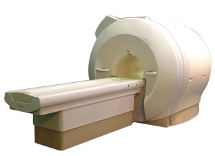
www.isoltech.co.kr/english/product/default.htm 'Next generation MRI system 1.5T CHORUS developed by ISOL Technology is optimized for both clinical diagnostic imaging and for research development.
CHORUS offers the complete range of feature oriented advanced imaging techniques- for both clinical routine and research. The compact short bore magnet, the patient friendly design and the gradient technology make the innovation to new degree of perfection in magnetic resonance.'
Device Information and Specification
CLINICAL APPLICATION
Whole body
CONFIGURATION
Short bore
Head, C-spine, L-spine, TMJ, Knee, Shoulder, General purpose, Phased Array System: 4 digital receiver channels (Up to 12 channels)
Optional
SYNCHRONIZATION
ECG/peripheral: Optional/yes, respiratory gating
PULSE SEQUENCES
Spin Echo, Gradient Echo, Fast Spin Echo, Inversion Recovery (STIR, Fluid Attenuated Inversion Recovery), FLASH, FISP, PSIF, Turbo Flash ( MPRAGE ),TOF MR Angiography, Standard echo planar imaging package (SE-EPI, GE-EPI), Optional: Advanced P.A. Imaging Package (up to 4 ch.), Advanced echo planar imaging package, Single Shot and Diffusion Weighted EPI, IR/FLAIR EPI
IMAGING MODES
2D/3D, Travelling Sat, Multi-Slab 3D, MTC and TONE Pulse Sequence, Fat/Water Suppression, Presaturation (up to 6 bands), Flow Compensation using GMR pulse, Multi-Slice, Multi-Group Imaging
SINGLE/MULTI SLICE
Image reconstruction time (2562 ) : 0.02 s
FOV
40 cm
BORE DIAMETER
or W x H
58 cm diameter (patient)
MAGNET WEIGHT
4050 kg
H*W*D
233 x 206 x 160 cm
COOLING SYSTEM TYPE
Water-cooled coil and air-cooled amplifier
CRYOGEN USE
0.07 L/hr helium
STRENGTH
20 mT/m (Upto 27 mT/m)
5-GAUSS FRINGE FIELD
2.5 m / 3.8 m
Passive and active
spacer

• View the DATABASE results for 'CHORUS 1.5T™' (2).Open this link in a new window

MRI Resources 
Absorption and Emission - Examinations - Societies - Cochlear Implant - Mobile MRI - Breast MRI
 
previous      51 - 55 (of 113)     next
Result Pages : [1 2 3]  [4 5 6 7 8 9 10 11 12 13 14 15 16 17 18 19 20 ... ]
 Random Page
 
Share This Page
FacebookTwitterLinkedIn

MR-TIP    
Community   
User
Pass
Forgot your UserID/Password ?    


Personalized protocols (age, gender, body habitus, etc.) lead to :
more automated planning 
improved patient comfort 
better diagnostics 
optimized image quality 
nothing 

Look
      Ups


MR-TIP.com uses cookies! By browsing MR-TIP.com, you agree to our use of cookies.

Magnetic Resonance - Technology Information Portal
Member of SoftWays' Medical Imaging Group - MR-TIP • Radiology-TIP • Medical-Ultrasound-Imaging • 
Copyright © 2003 - 2024 SoftWays. All rights reserved. [ 21 November 2024]
Terms of Use | Privacy Policy | Advertising
 [last update: 2024-02-26 03:41:00]