 | Info
Sheets |
| | | | | | | | | | | | | | | | | | | | | | | | |
 | Out-
side |
| | | | |
|
| | | | | |  | Searchterm 'Gauss' was also found in the following services: | | | | |
|  |  |
| |
|

If a device is to be labeled MR Safe, the following information should be provided:
•
Data demonstrating that when the device is introduced or used in the MRI environment (i.e. the MRI scan room) it does not pose an increased safety risk to the patient or other personnel,
•
a scientifically-based rationale for why data are not necessary to prove the safety of the device in the MR environment (for example, a passive device made entirely of a polymer known to be nonreactive in strong magnetic fields).
If a device is to be labeled MR Compatible, the following information should be provided:
•
Data demonstrating that when the device is introduced or used in the MRI environment, it is MR safe that it performs its intended function without performance degradation, and that it does not adversely affect the function of the MRI scanner (e.g. no significant image artifacts or noise). Any image artifact or noise due to the medical device should be quantified (e.g., % volume affected, signal to noise ratio),
•
a scientifically-based rationale for why data are not necessary to prove the compatibility of the device in the MRI environment.
Test Conditions:
The static magnetic field strength ( Gauss (G) or Tesla (T)) to which the device was tested and demonstrated to be MRI 'safe', 'compatible', or 'intended for use in' should be related to typical machine ratings (e.g. 0.5 T, 1.5 T, 2.0 T, and shielded or unshielded magnet, etc).
The same conditions should be used for the spatial gradient ( field strength per unit distance (i.e., G/cm)) in which the device was tested and demonstrated to be 'safe', 'compatible', or 'intended for use in'.
Also the RF transmitter power used during testing of the device, should be related to this typical machine ratings. | |  | | • For this and other aspects of MRI safety see our InfoSheet about MRI Safety. | | | • Patient-related information is collected in our MRI Patient Information.
| | |
• View the NEWS results for 'MR Compatibility' (2).
| | | | |  Further Reading: Further Reading: | | Basics:
|
|
News & More:
| |
| |
|  |  | Searchterm 'Gauss' was also found in the following service: | | | | |
|  |  |
| |
|
| |  | |
• View the DATABASE results for 'MR Guided Interventions' (8).
| | | | |  Further Reading: Further Reading: | Basics:
|
|
News & More:
|  |
AI analysis finds younger AFib patients benefit from MRI-guided ablation treatments
Friday, 25 August 2023 by www.eurekalert.org |  |  |
Theranostic nano-platform for MRI-guided synergistic therapy against breast cancer
Monday, 26 September 2022 by phys.org |  |  |
Magnetic seeds used to heat and kill cancer
Tuesday, 1 February 2022 by www.sciencedaily.com |  |  |
What is the effect of MRI with targeted biopsies on the rate of patients discontinuing active surveillance? A reflection of the use of MRI in the PRIAS study
Thursday, 8 April 2021 by www.docwirenews.com |  |  |
Modeling of Active Shimming of Metallic Needles for Interventional MRI
Monday, 29 June 2020 by pubmed.ncbi.nlm.nih.gov |  |  |
Magnetic Resonance Imaging Guided Confirmatory Biopsy for Initiating Active Surveillance of Prostate Cancer
Wednesday, 11 September 2019 by jamanetwork.com |  |  |
FDA clears ViewRay's next-gen, MRI-guided radiation therapy device
Tuesday, 28 February 2017 by www.fiercebiotech.com |  |  |
Siemens, U. of Twente Biopsy Robot Promises Greater Precision, Less Cost
Friday, 22 January 2016 by www.meddeviceonline.com |  |  |
Magnetic resonance-guided motorized transcranial ultrasound system for blood-brain barrier permeabilization along arbitrary trajectories in rodents
Thursday, 24 December 2015 by www.ncbi.nlm.nih.gov |  |  |
New MRI-Guided Catheter Shows Major Potential for Stroke Treatment
Tuesday, 29 December 2015 by www.radiology.ucsf.edu |  |  |
Polish study on MRI-ultrasound for targeted prostate biopsy wins CEM award
Tuesday, 12 November 2013 by medicalxpress.com |  |  |
C4 Imaging Announces FDA 510(k) Clearance of its Positive-Signal MRI Marker - Sirius™
Friday, 6 December 2013 by www.digitaljournal.com |
|
| |
|  |  | MRI Safety Resources | | | | |
|  |  |  |
| |
|

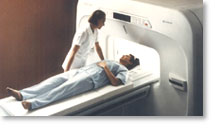
From Hitachi Medical Systems America, Inc.; because of its dependability, the MRP-7000™ remains popular more than a decade after the first U.S. system was shipped. This system maintains a high resale value, what has made it one of the most sought-after scanners on the used MRI equipment market.
Device Information and Specification CLINICAL APPLICATION Whole body DualQuad T/R Body Coil, MA Head, MA C-Spine, MA Shoulder, MA Wrist, MA CTL Spine, MA Knee, MA TMJ, MA Flex Body (3 sizes), Neck, small and large Extremity, PVA (WIP), Breast (WIP), Neurovascular (WIP), Cardiac (WIP) and MA Foot//Ankle (WIP) SE, GE, GR, IR, FIR, STIR, ss-FSE, FSE, DE-FSE/FIR, FLAIR, ss/ms-EPI, ss/ms EPI- DWI, SSP, MTC, SE/GE-EPI, MRCP, SARGE, RSSG, TRSG, BASG, Angiography: CE, PC, 2D/3D TOFIMAGING MODES Single, multislice, volume study horizontal 2.5 m x 2.1 m vertical | |  | |
• View the DATABASE results for 'MRP-7000™' (2).
| | | | |
|  |  | Searchterm 'Gauss' was also found in the following services: | | | | |
|  |  |
| |
|

From ONI Medical Systems, Inc.;
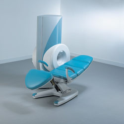
MSK-Extreme™ MRI system is a dedicated high field extremity imaging device, designed to provide orthopedic surgeons and other physicians with detailed diagnostic images of the foot, ankle, knee, hand, wrist and elbow, all with the clinical confidence and advantages derived from high field, whole body MRI units. The light weight (less than 650 kg) of the OrthOne System performs rapid patient studies, is easy to operate, has a patient friendly open environment and can be installed in a practice office or hospital, all at a cost similar to a low field extremity machine.
New features include a more powerful operating system that offers increased scan speed as well as a 160-mm knee coil with higher signal to noise ratio, and the option of a CD burner.
Device Information and Specification 16 cm knee, 18 cm lower extremity;; 12.3 cm upper extremity, additional high resolution v-SPEC Coils: 80 mm, 100 mm, or 145 mm. SE, FSE, GE2D, GE3D, Inversion recovery (IR), Driven Equilibrium, Fat Saturation (FS), STIR, MT, PD, Flow Compensation (FC), RF spoiling, MTE, No Phase Wrap (NPW) IMAGING MODES Scout, single, multislice, volume 2D less than 200 msec/image X/Y: 64-512; 2 pixel steps 4,096 grey lvls; 256 lvls in 3D POWER REQUIREMENTS 115VAC, 1phase, 20A; 208VAC, 3 phase, 30A COOLING SYSTEM TYPE LHe with 2 stage cold head 1.25m radial x 1.8m axial | |  | | | |  Further Reading: Further Reading: | Basics:
|
|
| |
|  |  | Searchterm 'Gauss' was also found in the following service: | | | | |
|  |  |
| |
|


O-scan is manufactured and distributed by Esaote SpA
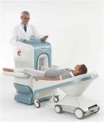
O-scan is a compact, dedicated extremity MRI system designed for easy installation and high throughput. The complete system fits in a 9' x 10' room, doesn't need for RF or magnetic shielding and it plugs in the wall. The 0.31T permanent magnet along with dual phased array RF coils, and advanced imaging protocols provide outstanding image quality and fast 25 minute complete examinations.
Esaote North America is the exclusive distributor of the O-scan system in the USA.
Device Information and Specification CLINICAL APPLICATION Dedicated Extremity
PULSE SEQUENCES
SE, HSE, HFE, GE, 2dGE, ME, IR, STIR, Stir T2, GESTIR, TSE, TME, FSE STIR, FSE ( T1, T2), X-Bone, Turbo 3DT1, 3D SHARC, 3D SST1, 3D SST2 2D: 2mm - 10 mm, 3D: 0.6 - 10 mm POWER REQUIREMENTS 100/110/200/220/230/240 | |  | | | |
|  | |  |  |
|  | | |
|
| |
 | Look
Ups |
| |