 | Info
Sheets |
| | | | | | | | | | | | | | | | | | | | | | | | |
 | Out-
side |
| | | | |
|
| | | | |
Result : Searchterm 'Gradient Strength' found in 1 term [ ] and 7 definitions [ ] and 7 definitions [ ], (+ 17 Boolean[ ], (+ 17 Boolean[ ] results ] results
| previous 6 - 10 (of 25) nextResult Pages :  [1] [1]  [2] [2]  [3 4 5] [3 4 5] |  | |  | Searchterm 'Gradient Strength' was also found in the following service: | | | | |
|  |  |
| |
|
(FOV) Defined as the size of the two or three dimensional spatial encoding area of the image. Usually defined in units of mm². The FOV is the square image area that contains the object of interest to be measured. The smaller the FOV, the higher the resolution and the smaller the voxel size but the lower the measured signal.
Useful for decreasing the scantime is a field of view different in the frequency and phase encoding directions ( rectangular field of view - RFOV).
The magnetic field homogeneity decreases as more tissue is imaged (greater FOV). As a result the precessional frequencies change across the imaging volume. That can be a problem for fat suppression imaging. This fat is precessing at the expected frequency only in the center of the imaging volume. E.g. frequency specific fat saturation pulses become less effective when the field of view is increased. It is best to use smaller field of views when applying fat saturation pulses.

Image Guidance
Smaller FOV required higher gradient strength and concludes low signal. Therefore you have to find a compromise between these factors.
The right choice of the field of view is important for MR image quality. When utilizing small field of views and scanning at a distance from the isocenter (more problems with artifacts) it is obviously important to ensure that the region of interest is within the scanning volume.
A smaller FOV in one direction is available with the function rectangular field of view (RFOV).
See also Field Inhomogeneity Artifact. | | | |  | | | | | | | | |  Further Reading: Further Reading: | | Basics:
|
|
News & More:
| |
| |
|  | |  |  |  |
| |
|
The owner of MRI equipment has to ensure that the equipment does fulfill the local requirements.
In some countries, the requirements are more stringent than in others; in other countries, they are nonexistent.
The user in general is unable to check power output, gradient strength, or even field strength.
The manufacturer has to cover authorized hardware and software updates after the initial installation and has to give guarantee for the requirements.
Specially designed computer programs usually supervise the power output of MRI devices and will not allow or will interrupt any imaging or spectroscopy procedure exceeding those limits considered safe.
See also European Medicines Agency, FDA information:
www.fda.gov/cdrh/safety/mrisafety.html | |  | |
• View the DATABASE results for 'Legal Requirements' (3).
| | | | |  Further Reading: Further Reading: | News & More:
|
|
| |
|  | |  |  | |  |  | Searchterm 'Gradient Strength' was also found in the following service: | | | | |
|  |  |
| |
|
Device Information and Specification
CLINICAL APPLICATION
Whole body
CONFIGURATION
Cylindrical Wide Short Bore
Opt. (WIP) Single and Multi Voxel
SE, FE, IR, FastSE, FastIR, FastFLAIR, Fast STIR, FastFE, FASE, Hybrid EPI, Multi Shot EPI; Angiography: 2D(gate/non-gate)/3D TOF, SORS-STC
IMAGING MODES
Single, multislice, volume study
TE
8 msec min. SE; 1.2 msec min. FE
less than 0.015 (256x256)
1.0 min. 2-DFT: 0.2 min. 3-DFT
32-1024, phase;; 64-1024, freq.
65.5 cm, patient aperture
4050 kg (bare magnet incl. L-He)
COOLING SYSTEM TYPE
Closed-loop water-cooled
Liquid helium: approx. less than 0.05 L/hr
Passive, active, auto-active
| |  | |
• View the DATABASE results for 'Excelart AG™ with Pianissimo' (2).
| | | | |
|  | |  |  |  |
| |
|

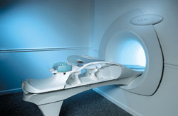
From Aurora Imaging Technology, Inc.;
The Aurora┬« 1.5T Dedicated Breast MRI System with Bilateral SpiralRODEO™ is the first and only FDA approved MRI device designed specifically for breast imaging. The Aurora System, which is already in clinical use at a growing number of leading breast care centers in the US, Europe, got in December 2006 also the approval from the State Food and Drug Administration of the People's Republic of China (SFDA).
'Some of the proprietary and distinguishing features of the Aurora System include: 1) an ellipsoid magnetic shim that provides coverage of both breasts, the chest wall and bilateral axillary lymph nodes; 2) a precision gradient coil with the high linearity required for high resolution spiral reconstruction;; 3) a patient-handling table that provides patient comfort and procedural utility; 4) a fully integrated Interventional System for MRI guided biopsy and localization; and 5) the user-friendly AuroraCADÔäó computer-aided image display system designed to improve the accuracy and efficiency of diagnostic interpretations.'
Device Information and Specification
CONFIGURATION
Short bore compact
TE
From 5 ms for RODEO Plus to over 80 ms, 120 ms for T2 sequences
Around 0.02 sec for a 256x256 image, 12.4 sec for a 512 x 512 x 32 multislice set
20 - 36 cm, max. elliptical 36 x 44 cm
POWER REQUIREMENTS
150A/120V-208Y/3 Phase//60 Hz/5 Wire
| |  | |
• View the DATABASE results for 'Aurora® 1.5T Dedicated Breast MRI System' (2).
| | |
• View the NEWS results for 'Aurora┬« 1.5T Dedicated Breast MRI System' (3).
| | | | |  Further Reading: Further Reading: | News & More:
|
|
| |
|  | |  |  |
|  | | |
|
| |
 | Look
Ups |
| |