Magnetic Resonance - Technology Information Portal Welcome to MRI Technology
Info
  Sheets

Out-
      side


 'H0' 
SEARCH FOR    
 
  2 3 5 A B C D E F G H I J K L M N O P Q R S T U V W X Y Z
Result : Searchterm 'H0' found in 1 term [] and 25 definitions []
1 - 5 (of 26)     next
Result Pages : [1]  [2 3 4 5 6]
MRI Resources 
RIS - Functional MRI - Contrast Enhanced MRI - Absorption and Emission - Homepages - IR
 
H0
 
[H0] Conventional symbol historically used for the constant magnetic field in an MR system; it is physically more correct to use B0.
spacer
 
• Share the entry 'H0':  Facebook  Twitter  LinkedIn  
MRI Resources 
Pregnancy - MRI Technician and Technologist Jobs - Research Labs - IR - Artifacts - Image Quality
 
B0
 
[B0] A conventional symbol for the main magnetic field strength (magnetic flux density or induction) in a MRI system. Although historically used, H0 (units of magnetic field strength, ampere//meter) should be distinguished from the more appropriate B0 [units of magnetic induction, tesla].
In current MR systems it has a constant value over time varying from 0.02 to 4 T. Field strengths of 0.5 T and above are generated with superconductive magnets. High field strengths have a better signal to noise ratio (SNR). The optimal imaging field strength for clinical imaging is between 0.5 and 2.0 T.

See also the related poll result: 'In 2010 your scanner will probably work with a field strength of'
spacer

• View the DATABASE results for 'B0' (41).Open this link in a new window

 
Further Reading:
  Basics:
Factors influencing flip angle mapping in MRI: RF pulse shape, slice-select gradients, off-resonance excitation, and B0 inhomogeneities.
Tuesday, 1 August 2006   by www.ncbi.nlm.nih.gov    
Magnetic Field
   by hyperphysics.phy-astr.gsu.edu    
  News & More:
Turbo-FLASH Based Arterial Spin Labeled Perfusion MRI at 7 T
Thursday, 20 June 2013   by www.plosone.org    
Penn researchers to get 7 Tesla whole-body MRI system
Monday, 28 August 2006   by www.eurekalert.org    
Optimizing SPIR and SPAIR fat suppression
Tuesday, 30 November 2004   by clinical.netforum.healthcare.philips.com    
MRI Resources 
Service and Support - Supplies - Anatomy - Breast Implant - Implant and Prosthesis pool - MRI Physics
 
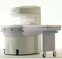
MAGNETOM Jazz™InfoSheet: - Devices -
Intro, 
Types of Magnets, 
Overview, 
etc.MRI Resource Directory:
 - Devices -
 
www.med.siemens.com/med/d/gg/mr/products/jazz.htm From Siemens Medical Systems;
This open MRI system is a dedicated extremity scanner, which is easy to use and easy to install. Whether you want to use it as a supplement to your whole-body scanner or you want to specialize in orthopedic exams, MAGNETOM Jazz™ offers you a cost effective opportunity to make the most of your capabilities.
Device Information and Specification
CLINICAL APPLICATION
CONFIGURATION
PULSE SEQUENCES
GRE, IR, FIR, STIR, TrueIR/FISP, FSE, MT, SS-FSE, MT-SE, MTC, MSE, GMR, fat/water sat./exc.
IMAGING MODES
Single, multislice, volume study, multi angle
Min 2D/3D: 0.1/0.05 mm
512 x 512 full screen display
MEASURING MATRIX
64 x 64 to 512 x 512
22 micrometer
MAGNET TYPE
Permanent
MAGNET WEIGHT
1800 kg
POWER REQUIREMENTS
380/400/420/440/480 V
STRENGTH
20 mT/m
Passive, active
spacer

• View the DATABASE results for 'MAGNETOM Jazz™' (2).Open this link in a new window

MRI Resources 
Hospitals - Examinations - Services and Supplies - Societies - Corporations - Fluorescence
 
MRP-7000™InfoSheet: - Devices -
Intro, 
Types of Magnets, 
Overview, 
etc.MRI Resource Directory:
 - Devices -
 
www.hitachimed.com/products/mrp.asp From Hitachi Medical Systems America, Inc.;
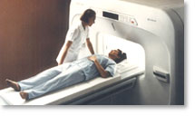
because of its dependability, the MRP-7000™ remains popular more than a decade after the first U.S. system was shipped. This system maintains a high resale value, what has made it one of the most sought-after scanners on the used MRI equipment market.
Device Information and Specification
CLINICAL APPLICATION
Whole body
CONFIGURATION
Vertical Orientation Field
DualQuad T/R Body Coil, MA Head, MA C-Spine, MA Shoulder, MA Wrist, MA CTL Spine, MA Knee, MA TMJ, MA Flex Body (3 sizes), Neck, small and large Extremity, PVA (WIP), Breast (WIP), Neurovascular (WIP), Cardiac (WIP) and MA Foot//Ankle (WIP)
SYNCHRONIZATION
Cardiac gating, ECG/peripheral, respiratory gating (2 modes)
PULSE SEQUENCES
SE, GE, GR, IR, FIR, STIR, ss-FSE, FSE, DE-FSE/FIR, FLAIR, ss/ms-EPI, ss/ms EPI- DWI, SSP, MTC, SE/GE-EPI, MRCP, SARGE, RSSG, TRSG, BASG, Angiography: CE, PC, 2D/3D TOF
IMAGING MODES
Single, multislice, volume study
MAGNET TYPE
Permanent, self shielded
STRENGTH
8 mT/m
5-GAUSS FRINGE FIELD
horizontal 2.5 m x 2.1 m vertical
Auto shimming, 3-axis/patient and active shimming
spacer

• View the DATABASE results for 'MRP-7000™' (2).Open this link in a new window

MRI Resources 
Safety pool - Open Directory Project - Equipment - PACS - Resources - Developers
 
MagneVu 1000InfoSheet: - Devices -
Intro, 
Types of Magnets, 
Overview, 
etc.MRI Resource Directory:
 - Devices -
 
www.mri4ra.com/isis.html From MagneVu;
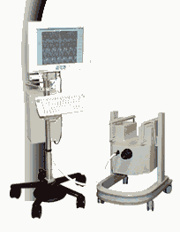
The MagneVu 1000 is a compact, robust, and portable, permanent magnet MRI system and operates without special shielding or costly site preparation.
This MRI device utilizes a patented non-homogeneous magnetic field image acquisition method to achieve high performance imaging. The MagneVu 1000 MRI scanner is designed for MRI of the extremities with the current specialty areas in diabetes and rheumatoid arthritis. Easy access is afforded for claustrophobic, pediatric, or limited mobility patients.
In August 1998 FDA marketing clearance and other regulatory approvals have been received.
Until 2008, over 130 devices in the US are in use. Some further developments of MagneVu's extremity scanner are: 'truly Plug n' Play MRI™' and iSiS ( which adds wireless capability to the second generation MV1000-XL).
Device Information and Specification
CLINICAL APPLICATION
Dedicated extremity
CONFIGURATION
Portable open MRI
IMAGING MODES
3-dimensional multi-echo data acquisition
3D: 0.6-1 mm
MAGNET TYPE
Permanent
MAGNET WEIGHT
about 50 kg
POWER REQUIREMENTS
110 V
spacer

• View the DATABASE results for 'MagneVu 1000' (3).Open this link in a new window

 
Further Reading:
  News & More:
VALUE OF 3D T1W & STIR MRI SEQUENCES IN DIAGNOSING EROSIONS IN RHEUMATOID ARTHRITIS
   by www.bocaradiology.com    
MRI Resources 
Absorption and Emission - MRI Physics - Databases - MRI Centers - Nerve Stimulator - Crystallography
 
     1 - 5 (of 26)     next
Result Pages : [1]  [2 3 4 5 6]
 Random Page
 
Share This Page
FacebookTwitterLinkedIn

MR-TIP    
Community   
User
Pass
Forgot your UserID/Password ?    


How AI will impact MRI :
only diagnostics 
saving time 
reducing cost 
makes planning obsolete 
reduce human knowledge 
not at all 

Look
      Ups


MR-TIP.com uses cookies! By browsing MR-TIP.com, you agree to our use of cookies.

Magnetic Resonance - Technology Information Portal
Member of SoftWays' Medical Imaging Group - MR-TIP • Radiology-TIP • Medical-Ultrasound-Imaging • 
Copyright © 2003 - 2024 SoftWays. All rights reserved. [ 28 January 2025]
Terms of Use | Privacy Policy | Advertising
 [last update: 2024-02-26 03:41:00]