Magnetic Resonance - Technology Information Portal Welcome to MRI Technology
Info
  Sheets

Out-
      side


 'Intera' 
SEARCH FOR    
 
  2 3 5 A B C D E F G H I J K L M N O P Q R S T U V W X Y Z
Result : Searchterm 'Intera' found in 9 terms [] and 36 definitions []
previous     21 - 25 (of 45)     next
Result Pages : [1 2]  [3 4 5 6 7 8 9]
Searchterm 'Intera' was also found in the following services: 
spacer
News  (9)  Resources  (22)  Forum  (10)  
 
DecouplingInfoSheet: - Artifacts - 
Case Studies, 
Reduction Index, 
etc.
 
1) The specific irradiation designed to remove the multiple structure in a particular resonance due to spin spin coupling with other nuclei;
2) The preventing of the interaction by mutual inductive coupling of two (or more) resonant RF coils, e.g. by detuning coils not in use at a particular point in time. Decoupling can take the form of active decoupling where an externally controlled switching circuit is used to detune the non-selected coils or passive decoupling where RF energy from the transmitter pulse is used to switch diodes to detune the appropriate coil.
spacer
 
Further Reading:
  Basics:
A Half-Volume Coil for Efficient Proton Decoupling in Humans at 4 Tesla
1997   by www.cmrr.umn.edu    
MRI Resources 
Musculoskeletal and Joint MRI - Cardiovascular Imaging - Pediatric and Fetal MRI - Devices - Collections - Breast MRI
 
Electron Spin Resonance
 
(ESR) Electron spin resonance is a spectroscopic technique to identify paramagnetic substances. This magnetic resonance phenomenon investigates the nature of the bonding within molecules by identifying unpaired electrons, e.g. in free radicals and their interaction with their immediate surroundings. The Larmor frequency are much higher than corresponding NMR frequencies in the same static magnetic field.
Nuclei with an odd number of neutrons and/or protons, because of their spin, react like tiny magnets and can be lined up in an applied magnetic field. Energy applied by alternating radio frequency radiation is absorbed when its frequency coincides with that of precession of the electron magnets. The spectrum of radiation absorbed as the field changes gives information valuable in chemistry, biology, and medicine since over 50 years.
spacer

• View the DATABASE results for 'Electron Spin Resonance' (2).Open this link in a new window


• View the NEWS results for 'Electron Spin Resonance' (1).Open this link in a new window.
 
Further Reading:
  Basics:
Electron Spin Resonance
   by hyperphysics.phy-astr.gsu.edu    
  News & More:
After merging resources, chemist, retina surgeon see melanin's value in new light
Thursday, 18 August 2005   by chronicle.uchicago.edu    
MRI Resources 
Devices - Resources - Mass Spectrometry - Anatomy - Veterinary MRI - DICOM
 
Fast Spin EchoForum -
related threadsInfoSheet: - Sequences - 
Intro, 
Overview, 
Types of, 
etc.
 
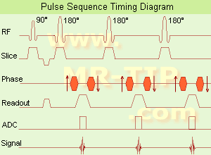
Fast Spin Echo Diagram (FSE) In the pulse sequence timing diagram, a fast spin echo sequence with an echo train length of 3 is illustrated. This sequence is characterized by a series of rapidly applied 180° rephasing pulses and multiple echoes, changing the phase encoding gradient for each echo.
The echo time TE may vary from echo to echo in the echo train. The echoes in the center of the K-space (in the case of linear k-space acquisition) mainly produce the type of image contrast, whereas the periphery of K-space determines the spatial resolution. For example, in the middle of K-space the late echoes of T2 weighted images are encoded. T1 or PD contrast is produced from the early echoes.
The benefit of this technique is that the scan duration with, e.g. a turbo spin echo turbo factor / echo train length of 9, is one ninth of the time. In T1 weighted and proton density weighted sequences, there is a limit to how large the ETL can be (e.g. a usual ETL for T1 weighted images is between 3 and 7). The use of large echo train lengths with short TE results in blurring and loss of contrast. For this reason, T2 weighted imaging profits most from this technique.
In T2 weighted FSE images, both water and fat are hyperintense. This is because the succession of 180° RF pulses reduces the spin spin interactions in fat and increases its T2 decay time. Fast spin echo (FSE) sequences have replaced conventional T2 weighted spin echo sequences for most clinical applications. Fast spin echo allows reduced acquisition times and enables T2 weighted breath hold imaging, e.g. for applications in the upper abdomen.
In case of the acquisition of 2 echoes this type of a sequence is named double fast spin echo / dual echo sequence, the first echo is usually density and the second echo is T2 weighted image. Fast spin echo images are more T2 weighted, which makes it difficult to obtain true proton density weighted images. For dual echo imaging with density weighting, the TR should be kept between 2000 - 2400 msec with a short ETL (e.g., 4).
Other terms for this technique are:
Turbo Spin Echo
Rapid Imaging Spin Echo,
Rapid Spin Echo,
Rapid Acquisition Spin Echo,
Rapid Acquisition with Refocused Echoes
 
Images, Movies, Sliders:
 Lumbar Spine T2 FSE Sagittal  Open this link in a new window
    

Courtesy of  Robert R. Edelman
 MRI - Anatomic Imaging of the Foot  Open this link in a new window
    
SlidersSliders Overview

 Lumbar Spine T2 FSE Axial  Open this link in a new window
    

Courtesy of  Robert R. Edelman
 
spacer

• View the DATABASE results for 'Fast Spin Echo' (31).Open this link in a new window

 
Further Reading:
  Basics:
MYELIN-SELECTIVE MRI: PULSE SEQUENCE DESIGN AND OPTIMIZATION
   by www.imaging.robarts.ca    
Advances in Magnetic Resonance Neuroimaging
Friday, 27 February 2009   by www.ncbi.nlm.nih.gov    
  News & More:
New MR sequence helps radiologists more accurately evaluate abnormalities of the uterus and ovaries
Thursday, 23 April 2009   by www.eurekalert.org    
Spin echoes, CPMG and T2 relaxation - Introductory NMR & MRI from Magritek
2013   by www.azom.com    
Searchterm 'Intera' was also found in the following services: 
spacer
News  (9)  Resources  (22)  Forum  (10)  
 
Functional Magnetic Resonance ImagingMRI Resource Directory:
 - Functional MRI -
 
(fMRI) Functional magnetic resonance imaging is a technique used to determine the dynamic brain function, often based on echo planar imaging, but can also be performed by using contrast agents and observing their first pass effects through brain tissue. Functional magnetic resonance imaging allows insights in a dysfunctional brain as well as into the basic workings of the brain.
The in functional brain MRI most frequently used effect to assess brain function is the blood oxygenation level dependent contrast (BOLD) effect, in which differential changes in brain perfusion and their resultant effect on the regional distribution of oxy- to deoxyhaemoglobin are observable because of the different 'intrinsic contrast media' effects of the two haemoglobin forms. Increased brain activity causes an increased demand for oxygen, and the vascular system actually overcompensates for this, increasing the amount of oxygenated haemoglobin. Because deoxygenated haemoglobin attenuates the MR signal, the vascular response leads to a signal increase that is related to the neural activity.
Functional imaging relates body function or thought to specific locations where the neural activity is taking place. The brain is scanned at low resolution but at a fast rate (typically once every 2-3 seconds). Structural MRI together with fMRI provides an anatomical baseline and best spatial resolution.
Interactions can also be seen from the motor cortex to the cerebellum or basal ganglia in the case of a movement disorder such as ataxia. For example: by a finger movement the briefly increase in the blood circulation of the appropriate part of the brain controlling that movement, can be measured.
spacer

• View the DATABASE results for 'Functional Magnetic Resonance Imaging' (8).Open this link in a new window


• View the NEWS results for 'Functional Magnetic Resonance Imaging' (15).Open this link in a new window.
 
Further Reading:
  Basics:
IMAGE CONTRAST IN MRI(.pdf)
   by www.assaftal.com    
  News & More:
New AI application reads eye movements
Tuesday, 16 November 2021   by www.news-medical.net    
Functional MRI Can Provide Clearer Picture of Unresponsive COVID-19 Patient's Brain Function and Potential for Recovery
Tuesday, 26 January 2021   by www.hospimedica.com    
Scientists first studied the brain of birds while moving
Sunday, 20 September 2020   by freenews.live    
MRI Technique Used to Identify Future Risk of Binge Drinking
Monday, 6 January 2020   by www.diagnosticimaging.com    
Functional MRI may help identify new, effective painkillers for chronic pain sufferers
Thursday, 4 February 2016   by www.eurekalert.org    
Study shows functional MRI differences in working memory in people with primary insomnia
Saturday, 31 August 2013   by www.news-medical.net    
Functional magnetic resonance imaging may improve diagnosis of autism
Tuesday, 31 May 2011   by www.dnaindia.com    
Using fMRI to study brain development
Friday, 30 November 2007   by www.eurekalert.org    
MRI Resources 
Case Studies - Universities - Manufacturers - Used and Refurbished MRI Equipment - Fluorescence - Crystallography
 
Gadopentetate DimeglumineInfoSheet: - Contrast Agents - 
Intro, Overview, 
Characteristics, 
Types of, 
etc.MRI Resource Directory:
 - Contrast Agents -
 
Short name: Gd-DTPA, generic name: Gadopentetate dimeglumine, chemical compound: Gadolinium-diethylenetriaminepentaacetic acid
Gadopentetate dimeglumine was introduced in 1981, as the first paramagnetic MRI contrast agent (ionic). The Gd-induced dipole dipole interactions lead to shortening of T1, which results in contrast enhancement on T1 weighted images. The used metal ion Gd3+ (gadolinium) is toxic, and therefore bound in the renally excreted DTPA chelate, a very stable complex. The Gd-complex also induce susceptibility effects, as a result of the magnetic field gradient between the contrast agent in the blood vessels and the surrounding tissue, that lead to shortening of T2 or T2*.
Following intravenous administration, the compound is distributed rapidly in the extracellular space and is eliminated unchanged by glomerular filtration via the kidneys. Up to 6 hours, post injection an average of 83% of the dose is eliminated renal.

See also Magnevist®, Gadolinium and Contrast Agents.
spacer

• View the DATABASE results for 'Gadopentetate Dimeglumine' (5).Open this link in a new window

 
Further Reading:
  Basics:
Magnevist Package Insert
2000
Gadopentetic acid
   by en.wikipedia.org    
  News & More:
EMA's final opinion confirms restrictions on use of linear gadolinium agents in body scans
Friday, 21 July 2017   by www.ema.europa.eu    
MRI Resources 
Research Labs - Distributors - Mobile MRI Rental - Universities - Collections - Intraoperative MRI
 
previous      21 - 25 (of 45)     next
Result Pages : [1 2]  [3 4 5 6 7 8 9]
 Random Page
 
Share This Page
FacebookTwitterLinkedIn

MR-TIP    
Community   
User
Pass
Forgot your UserID/Password ?    


MRI is trending to low field magnets :
reduced costs will lead to this change 
AI will close the gap to high field 
only in remote areas 
is only temporary 
never 

Look
      Ups


MR-TIP.com uses cookies! By browsing MR-TIP.com, you agree to our use of cookies.

Magnetic Resonance - Technology Information Portal
Member of SoftWays' Medical Imaging Group - MR-TIP • Radiology-TIP • Medical-Ultrasound-Imaging • 
Copyright © 2003 - 2024 SoftWays. All rights reserved. [ 21 November 2024]
Terms of Use | Privacy Policy | Advertising
 [last update: 2024-02-26 03:41:00]