 | Info
Sheets |
| | | | | | | | | | | | | | | | | | | | | | | | |
 | Out-
side |
| | | | |
|
| | | | |
Result : Searchterm 'Intervention' found in 1 term [ ] and 20 definitions [ ] and 20 definitions [ ] ]
| previous 11 - 15 (of 21) nextResult Pages :  [1] [1]  [2 3 4 5] [2 3 4 5] |  | |  | Searchterm 'Intervention' was also found in the following services: | | | | |
|  |  |
| |
|
Ultrasound imaging is the primary fetal monitoring modality during pregnancy, nevertheless fetal MRI is increasingly used to image anatomical regions and structures difficult to see with sonography. Given its long record of safety, utility, and cost-effectiveness, ultrasound will remain the modality of first choice in fetal screening. However, MRI is beginning to fill a niche in situations where ultrasound does not provide enough information to diagnose abnormalities before the baby's birth. Magnetic resonance imaging of the fetus provides multiplanar views also in sub-optimal positions, better characterization of anatomic details of e.g. the fetal brain, and information for planning the mode of delivery and airway management at birth.
Indications:
•
Examinations of the placenta
Modern fetal MRI requires no sedatives or muscle relaxants to control fetal movement. Ultrafast MRI techniques (e.g., single shot techniques like Half Fourier Acquisition Single shot Turbo spin Echo HASTE) enable images to be acquired in less than one second to eliminate fetal motion. Such technology has led to increased usage of fetal MRI, which can lead to earlier diagnosis of conditions affecting the baby and has proven useful in planning fetal surgery and designing postnatal treatments. As MR technology continues to improve, more advances in the prenatal diagnosis and treatment of fetal abnormalities are to expect. More advances in in-utero interventions are likely as well. Eventually, fetal MRI may replace even some prenatal tests that require invasive procedures such as amniocentesis.
For Ultrasound Imaging (USI) see Fetal Ultrasound at Medical-Ultrasound-Imaging.com. | | | |  | | | | | | | | |  Further Reading: Further Reading: | | Basics:
|
|
News & More:
|  |
Advances in medical imaging enable visualization of white matter tracts in fetuses
Wednesday, 12 May 2021 by www.eurekalert.or |  |  |
Fetal CMR Detects Congenital Heart Defects, Changes Treatment Decisions
Monday, 29 March 2021 by www.diagnosticimaging.com |  |  |
MRI scans more precisely define and detect some abnormalities in unborn babies
Friday, 12 March 2021 by www.eurekalert.org |  |  |
Ultrasound and Magnetic Resonance Imaging of Agenesis of the Corpus Callosum in Fetuses: Frontal Horns and Cavum Septi Pellucidi Are Clues to Earlier Diagnosis
Monday, 29 June 2020 by pubmed.ncbi.nlm.nih.gov |  |  |
MRI helps predict preterm birth
Tuesday, 15 March 2016 by www.eurekalert.org |  |  |
3-T MRI advancing on ultrasound for imaging fetal abnormalities
Monday, 20 April 2015 by www.eurekalert.org |  |  |
Babies benefit from pioneering 'miniature' MRI scanner in Sheffield
Friday, 24 January 2014 by www.telegraph.co.uk |  |  |
Ultrasensitive Detector Pinpoints Big Problem in Tiny Fetal Heart
Tuesday, 6 April 2010 by www.sciencedaily.com |  |  |
Real-time MRI helps doctors assess beating heart in fetus
Thursday, 29 September 2005 by www.eurekalert.org |
|
| |
|  | |  |  |  |
| |
|

GE Healthcare is the result of the merger between GE Medical and Amersham Health in Nov. 2004, after GE acquired Amersham Health for 9.5 billion in Oct. 2003. Jeffrey R. Immelt, Chairman of the Board and Chief Executive of General Electric, said, 'Amersham's diagnostic pharmaceutical and life sciences business will add new, high growth platforms to GE Medical's diagnostic imaging, services and healthcare information technology businesses'. GE Healthcare, a UK company, is a unit of General Electric (NYSE: GE). GE Healthcare is a global leader in medical imaging, diagnostic imaging contrast agents, interventional procedures, healthcare services, and information technology.
For more than 100 years, health care providers have relied on GE Medical Systems, now GE Healthcare, for high quality medical technology and productivity solutions.
GE Healthcare, headquartered now at formerly seat of Amersham Health in Great Britain, operates facilities around the world. Global Operations include organizations on the Americas, Europe, and Asia, including India, Japan, Korea China, Thailand and Vietnam.
MRI Scanners:
0.2T to 1.0T:
to 1.5T:
to 3.0T:
MRI Contrast Agents:
| |  | |
• View the DATABASE results for 'GE Healthcare' (23).
| | |
• View the NEWS results for 'GE Healthcare' (26).
| | | | |  Further Reading: Further Reading: | Basics:
|
|
News & More:
| |
| |
|  | |  |  |  |
| |
|

From Philips Medical Systems;
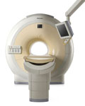
The clinical capabilities of MR will further expand. Inside and out, the Achieva is a friendly, open system designed for optimal patient comfort and maximized workflow with high functionality.
The Achieva 1.5T can be upgraded to Achieva I/T, with three configurations optimized for MR guided interventions and therapy:
•
Achieva I/T Neurosurgery
•
Achieva I/T Cardiovascular (or XMR - combining an Achieva 1.5T CV system and an X-Ray system)
Device Information and Specification
CLINICAL APPLICATION
Whole body
CONFIGURATION
Short bore compact
Standard: Head, body, C1, C3; Optional: Small joint, flex-E, flex-R, endocavitary (L and S), dual TMJ, knee, neck, T/L spine, breast; optional phased array: Spine, pediatric, 3rd party connector; Optional SENSEâ„¢ coils for all applications
SE, Modified-SE, IR (T1, T2, PD), STIR, FLAIR, SPIR, FFE, T1-FFE, T2-FFE, Balanced FFE, TFE, Balanced TFE, Dynamic, Keyhole, 3D, Multi Chunk 3D, Multi Stack 3D, K Space Shutter, MTC, TSE, Dual IR, DRIVE, EPI, Cine, 2DMSS, DAVE, Mixed Mode; Angiography: Inflow MRA, TONE, PCA, CE MRA
128 x 128, 256 x 256,512 x 512,1024 x 1024 (64 for Bold img)
Variable in 1% increments
Lum.: 120 cd/m2; contrast: 150:1
Variable (op. param. depend.)
POWER REQUIREMENTS
380/400 V
| |  | |
• View the DATABASE results for 'Intera Achieva 1.5T™' (2).
| | | | |
|  |  | Searchterm 'Intervention' was also found in the following services: | | | | |
|  |  |
| |
|
With an open configuration MRI system neurosurgical procedures can be performed using image guidance. Open MRI can be used to guide interventional treatments or procedures, such as a biopsy.
Intraoperative MRI allows lesions to be precisely localized and targeted.
Constantly updated images, correlated with images obtained pre-operatively, help to eliminate errors that can arise during framed and frameless stereotactic surgery when anatomic structures alter their position due to shifting or displacement of, e.g. brain parenchyma. Intraoperative MRI can help with the identification of normal structures, such as blood vessels and is helpful in optimizing surgical approaches, achieving complete resection of intracerebral lesions, determining tumor margins and monitoring potential intraoperative complications. | |  | |
• View the DATABASE results for 'Intraoperative Magnetic Resonance Imaging' (4).
| | |
• View the NEWS results for 'Intraoperative Magnetic Resonance Imaging' (1).
| | | | |  Further Reading: Further Reading: | Basics:
|
|
News & More:
| |
| |
|  | |  |  |  |
| |
|
| |  | |
• View the DATABASE results for 'Low Field MRI' (8).
| | |
• View the NEWS results for 'Low Field MRI' (5).
| | | | |  Further Reading: Further Reading: | Basics:
|
|
News & More:
|  |
Safety of Bedside Portable Low-Field Brain MRI in ECMO Patients Supported on Intra-Aortic Balloon Pump
Friday, 18 November 2022 by www.mdpi.com |  |  |
Researchers at the University of Tsukuba develop a portable MRI system specifically for identifying wrist cartilage damage among athletes, providing a convenient means of early detection and treatment of injuries
Tuesday, 26 April 2022 by www.tsukuba.ac.jp |  |  |
This bizarre looking helmet can create better brain scans
Friday, 11 February 2022 by www.sciencedaily.com |  |  |
A low-cost and shielding-free ultra-low-field brain MRI scanner
Tuesday, 14 December 2021 by www.nature.com |  |  |
Portable MRI provides life-saving information to doctors treating strokes
Thursday, 5 August 2021 by news.yale.edu |  |  |
Synaptive Evry, an MRI for Any Space, Cleared by FDA
Thursday, 30 April 2020 by www.medgadget.com |  |  |
World's First Portable MRI Cleared by FDA
Monday, 17 February 2020 by www.medgadget.com |  |  |
Introducing a point-of-care MRI system
Tuesday, 29 October 2019 by healthcare-in-europe.com |  |  |
Opportunities in Interventional and Diagnostic Imaging by Using High-performance Low-Field-Strength MRI
Tuesday, 1 October 2019 by pubs.rsna.org |  |  |
Portable 'battlefield MRI' comes out of the lab
Thursday, 30 April 2015 by physicsworld.com |  |  |
Portable MRI could aid wounded soldiers and children in the third world
Thursday, 23 April 2015 by phys.org |
|
| |
|  | |  |  |
|  | | |
|
| |
 | Look
Ups |
| |