 | Info
Sheets |
| | | | | | | | | | | | | | | | | | | | | | | | |
 | Out-
side |
| | | | |
|
| | | | |
Result : Searchterm 'Knee MRI' found in 1 term [ ] and 3 definitions [ ] and 3 definitions [ ], (+ 19 Boolean[ ], (+ 19 Boolean[ ] results ] results
| 1 - 5 (of 23) nextResult Pages :  [1] [1]  [2 3 4 5] [2 3 4 5] |  | |  | Searchterm 'Knee MRI' was also found in the following services: | | | | |
|  |  |
| |
|
Knee MRI, with its high soft tissue contrast is one of the main imaging tools to depict knee joint pathology. MRI allows accurate imaging of intra-articular structures such as ligaments, cartilage, menisci, bone marrow, synovium, and adjacent soft tissue.
Knee exams require a dedicated extremity coil, providing a homogenous imaging volume and high SNR to ensure best signal coverage.
A complete knee MR examination includes for example sagittal and coronal T1 weighted, and proton density weighted pulse sequences +/- fat saturation, or STIR sequences. For high spatial resolution, maximal 4 mm thick slices with at least an in plane resolution of 0.75 mm and small gap are recommended. To depict the anterior cruciate ligament clearly, the sagittal plane has to be rotated 10 - 20° externally (parallel to the medial border of the femoral condyle). Retropatellar cartilage can bee seen for example in axial T2 weighted gradient echo sequences with Fatsat. However, the choice of the pulse sequences is depended of the diagnostic question, the used scanner, and preference of the operator.
Diagnostic quality in knee imaging is possible with field strengths ranging from 0.2 to 3T. With low field strengths more signal averages must be measured, resulting in increased scan times to provide equivalent quality as high field strengths.
More diagnostic information of meniscal tears and chondral defects can be obtained by direct magnetic resonance arthrography, which is done by introducing a dilute solution of gadolinium in saline (1:1000) into the joint capsule. The knee is then scanned in all three planes using T1W sequences with fat suppression. For indirect arthrography, the contrast is given i.v. and similar scans are started 20 min. after injection and exercise of the knee.
Frequent indications of MRI scans in musculoskeletal knee diseases are: e.g., meniscal degeneration and tears, ligament injuries, osteochondral fractures, osteochondritis dissecans, avascular bone necrosis and rheumatoid arthritis. See also Imaging of the Extremities and STIR. | | | |  | | | | • Share the entry 'Knee MRI':    | | | | | | | | | |  Further Reading: Further Reading: | | Basics:
|
|
News & More:
| |
| |
|  |  | Searchterm 'Knee MRI' was also found in the following service: | | | | |
|  |  |
| |
|
Knee and shoulder MRI exams are the most commonly requested musculoskeletal MRI scans. Other MR imaging of the extremities includes hips, ankles, elbows, and wrists. Orthopedic imaging requires very high spatial resolution for reliable small structure definition and therefore places extremely high demands on SNR.
Exact presentation of joint pathology expects robust and reliable fat suppression, often under difficult conditions like off-center FOV,
imaging at the edge of the field homogeneity or in regions with complex magnetic susceptibility.
MR examinations can evaluate meniscal dislocations, muscle fiber tears, tendon disruptions, tendinitis, and diagnose bone tumors and soft tissue masses. MR can also demonstrate acute fractures that are radiographically impossible to see. Evaluation of articular cartilage for traumatic injury or assessment of degenerative disease represents an imaging challenge, which can be overcome by high field MRI applications. Currently, fat-suppressed 3D spoiled gradient echo sequences and density weighted fast spin echo sequences are the gold-standard techniques used to assess articular cartilage.
Open MRI procedures allow the kinematic imaging of joints, which provides added value to any musculoskeletal MRI practice. This technique demonstrates the actual functional impingements or positional subluxations of joints. In knee MRI examinations, the kinematical patellar study can show patellofemoral joint abnormalities.
See also Open MRI, Knee MRI, Low Field MRI. | | | | | | | | | | |
• View the DATABASE results for 'Imaging of the Extremities' (5).
| | | | |  Further Reading: Further Reading: | Basics:
|
|
News & More:
| |
| |
|  | |  |  |  |
| |
|
Fat suppression is the process of utilizing specific MRI parameters to remove the deleterious effects of fat from the resulting images , e.g. with STIR, FAT SAT sequences, water selective (PROSET WATS - water only selection, also FATS - fat only selection possible) excitation techniques, or pulse sequences based on the Dixon method.
Spin magnetization can be modulated by using special RF pulses. CHESS or its variations like SPIR, SPAIR ( Spectral Selection Attenuated Inversion Recovery) and FAT SAT use frequency selective excitation pulses, which produce fat saturation.
Fat suppression techniques are nearly used in all body parts and belong to every standard MRI protocol of joints like knee, shoulder, hips, etc.

Image Guidance
Imaging of, e.g. the foot can induce bad fat suppression with SPIR/FAT SAT due to the asymmetric volume of this body part. The volume of the foot alters the magnetic field to a different degree than the smaller volume of the lower leg affecting the protons there. There is only a small band of tissue where the fat protons are precessing at the frequency expected, resulting in frequency selective fat saturation working only in that area. This can be corrected by volume shimming or creating a more symmetrical volume being imaged with water bags.
Even with their longer scan time and motion sensitivity, STIR (short T1/tau inversion recovery) sequences are often the better choice to suppress fat. STIR images are also preferred because of the decreased sensitivity to field inhomogeneities, permitting larger fields of views when compared to fat suppressed images and the ability to image away from the isocenter. See also Knee MRI.
Sequences based on Dixon turbo spin echo ( fast spin echo) can deliver a significant better fat suppression than conventional TSE/FSE imaging.
| | | |  | |
• View the DATABASE results for 'Fat Suppression' (28).
| | | | |  Further Reading: Further Reading: | Basics:
|
|
News & More:
| |
| |
|  |  | Searchterm 'Knee MRI' was also found in the following services: | | | | |
|  |  |
| |
|
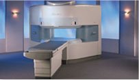
Open MRI scanners have been developed for people who are anxious or obese or for examination of small parts of the body, such as the extremities ( knee, shoulder). In addition, some systems offer imaging in different positions and sequences of movements.
The basic technology of an open MRI machine is similar to that of a traditional MRI device.
The major difference for the patient is that instead of lying in a narrow tunnel, the imaging table has more space around the body so that the magnet does not completely surround the person being tested.
Types of constructions:
•
Semi open high field MRI scanners provide an ultra short bore (tunnel) and widely flared ends. In this type of MRI systems, patients lie with the head in the space outside the bore, if for example the hips are examined.
•
Open low field MRI machines have often a wide open design, e.g. an open C-arm scanner is shaped like two large discs separated by a large pillar. Patients have an open sided feeling and more space around them allows a wider range of positions.
•
Advanced open MRI scanners combine the advantages of both, the high field strength, newest gradient technology and wide open design. Even scans of patients in upright, weight-bearing positions are possible (e.g. Upright™ MRI formerly Stand-Up MRI).
Difficulties with a traditional MRI scan include claustrophobia and patient size or, for health related reasons, patients who are not able to receive this type of diagnostic test. The MRI unit is a limited space, and some patients may be too large to fit in a narrow tunnel. In addition, weight limits can restrict the use of some scanners. The open MRI magnet has become the best option for those patients.
All of the highest resolution MRI scanners are tunnels and tend to accentuate the claustrophobic reaction. While patients may find the open MRI scanners easier to tolerate, some machines use a lower field magnet and generates lower image quality or have longer scan time. The better performance of an advanced open MRI scanner allows good image quality caused by the higher signal to noise ratio with maximum patient comfort.
See also Claustrophobia, MRI scan and Knee MRI. | |  | |
• View the DATABASE results for 'Open MRI' (37).
| | |
• View the NEWS results for 'Open MRI' (16).
| | | | |  Further Reading: Further Reading: | Basics:
|
|
News & More:
| |
| |
|  |  | Searchterm 'Knee MRI' was also found in the following service: | | | | |
|  |  |
| |
|

From Hitachi Medical Systems America, Inc.;
the AIRIS made its debut in 1995. Hitachi followed up with the AIRIS II system, which has proven equally successfully. 'All told, Hitachi has installed more than 1,000 MRI systems in the U.S., holding more than 17 percent of the total U.S. MRI installed base, and more than half of the installed base of open MR systems,' says Antonio Garcia, Frost and Sullivan industry research analyst.
Now Altaire employs a blend of innovative Hitachi features called VOSI™ technology, optimizing each sub-system's performance in concert with the
other sub-systems, to give the seamless mix of high-field performance
and the patient comfort, especially for claustrophobic patients, of open MR systems.
Device Information and Specification
CLINICAL APPLICATION
Whole body
DualQuad T/R Body Coil, MA Head, MA C-Spine, MA Shoulder, MA Wrist, MA CTL Spine, MA Knee, MA TMJ, MA Flex Body (3 sizes), Neck, small and large Extremity, PVA (WIP), Breast (WIP), Neurovascular (WIP), Cardiac (WIP) and MA Foot//Ankle (WIP)
SE, GE, GR, IR, FIR, STIR, ss-FSE, FSE, DE-FSE/FIR, FLAIR, ss/ms-EPI, ss/ms EPI- DWI, SSP, MTC, SE/GE-EPI, MRCP, SARGE, RSSG, TRSG, BASG, Angiography: CE, PC, 2D/3D TOF
IMAGING MODES
Single, multislice, volume study
TR
SE: 30 - 10,000msec GE: 3.6 - 10,000msec IR: 50 - 16,700msec FSE: 200 - 16,7000msec
TE
SE : 8 - 250msec IR: 5.2 -7,680msec GE: 1.8 - 2,000 msec FSE: 5.2 - 7,680
0.05 sec/image (256 x 256)
2D: 2 - 100 mm; 3D: 0.5 - 5 mm
Level Range: -2,000 to +4,000
COOLING SYSTEM TYPE
Water-cooled
3.1 m lateral, 3.6 m vertical
| |  | |
• View the DATABASE results for 'Altaire™' (2).
| | | | |  Further Reading: Further Reading: | News & More:
|
|
| |
|  | |  |  |
|  | 1 - 5 (of 23) nextResult Pages :  [1] [1]  [2 3 4 5] [2 3 4 5] |
| |
|
| |
 | Look
Ups |
| |