Magnetic Resonance - Technology Information Portal Welcome to MRI Technology
Info
  Sheets

Out-
      side


 'Line Width' 
SEARCH FOR    
 
  2 3 5 A B C D E F G H I J K L M N O P Q R S T U V W X Y Z
Result : Searchterm 'Line Width' found in 1 term [] and 3 definitions [], (+ 7 Boolean[] results
1 - 5 (of 11)     next
Result Pages : [1]  [2 3]
MRI Resources 
Services and Supplies - Intraoperative MRI - Spine MRI - Libraries - Fluorescence - Research Labs
 
Line Width
 
Spread in frequency of a resonance line in a MR spectrum. A common measure of the line width is full width at half maximum (FWHM).
spacer
 
• Share the entry 'Line Width':  Facebook  Twitter  LinkedIn  
 
Further Reading:
  Basics:
Full width at half maximum
   by en.wikipedia.org    
MRI Resources 
Shoulder MRI - Intraoperative MRI - Most Wanted - MRI Technician and Technologist Career - Homepages - RIS
 
Exponential Weighting
 
Multiplication of the time-dependent signal data by an exponential function, exp(t/TC), where t is time and TC is a parameter called the time constant (in spectroscopy). The time constant can be chosen to either improve the signal to noise ratio (with a negative TC) or decrease the effective spectral line width (with a positive TC) in the resulting spectrum. The use of a negative TC to improve SNR is equivalent to line broadening by convolving the spectrum with a Lorentzian function of corresponding reciprocal width.
spacer

• View the DATABASE results for 'Exponential Weighting' (2).Open this link in a new window

MRI Resources 
Anatomy - Used and Refurbished MRI Equipment - Open Directory Project - Breast Implant - RIS - MRCP
 
Gaussian Line Shape
 
A line shape characterized by a bell-shaped form; proportional to exp (-(f - f0)2/Df2) where Df is a measure of the line width.
spacer

• View the DATABASE results for 'Gaussian Line Shape' (2).Open this link in a new window

MRI Resources 
Services and Supplies - Intraoperative MRI - Jobs pool - Mass Spectrometry - Implant and Prosthesis pool - Calculation
 
Projection Profile
 
Spectrum of NMR signal whose frequency components are broadened by a magnetic field gradient. In the simplest case (negligible line width, no relaxation effects, and no effects of prior gradients), it corresponds to a one-dimensional projection of the spin density along the direction of the gradient; in this form it is used in projection reconstruction imaging.
spacer

• View the DATABASE results for 'Projection Profile' (4).Open this link in a new window

MRI Resources 
MRI Training Courses - Implant and Prosthesis - Distributors - Directories - MRI Technician and Technologist Jobs - Absorption and Emission
 
Echo Planar ImagingInfoSheet: - Sequences - 
Intro, 
Overview, 
Types of, 
etc.MRI Resource Directory:
 - Sequences -
 
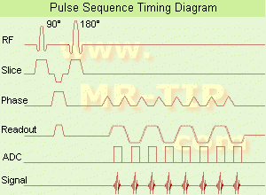
Echo Planar Imaging Timing Diagram (EPI) Echo planar imaging is one of the early magnetic resonance imaging sequences (also known as Intascan), used in applications like diffusion, perfusion, and functional magnetic resonance imaging. Other sequences acquire one k-space line at each phase encoding step. When the echo planar imaging acquisition strategy is used, the complete image is formed from a single data sample (all k-space lines are measured in one repetition time) of a gradient echo or spin echo sequence (see single shot technique) with an acquisition time of about 20 to 100 ms. The pulse sequence timing diagram illustrates an echo planar imaging sequence from spin echo type with eight echo train pulses. (See also Pulse Sequence Timing Diagram, for a description of the components.)
In case of a gradient echo based EPI sequence the initial part is very similar to a standard gradient echo sequence. By periodically fast reversing the readout or frequency encoding gradient, a train of echoes is generated.
EPI requires higher performance from the MRI scanner like much larger gradient amplitudes. The scan time is dependent on the spatial resolution required, the strength of the applied gradient fields and the time the machine needs to ramp the gradients.
In EPI, there is water fat shift in the phase encoding direction due to phase accumulations. To minimize water fat shift (WFS) in the phase direction fat suppression and a wide bandwidth (BW) are selected. On a typical EPI sequence, there is virtually no time at all for the flat top of the gradient waveform. The problem is solved by "ramp sampling" through most of the rise and fall time to improve image resolution.
The benefits of the fast imaging time are not without cost. EPI is relatively demanding on the scanner hardware, in particular on gradient strengths, gradient switching times, and receiver bandwidth. In addition, EPI is extremely sensitive to image artifacts and distortions.
spacer

• View the DATABASE results for 'Echo Planar Imaging' (19).Open this link in a new window


• View the NEWS results for 'Echo Planar Imaging' (1).Open this link in a new window.
 
Further Reading:
  Basics:
New Imaging Method Makes Brain Scans 7 Times Faster
Sunday, 9 January 2011   by www.dailytech.com    
MRI Resources 
Most Wanted - Crystallography - Societies - Service and Support - Intraoperative MRI - PACS
 
     1 - 5 (of 11)     next
Result Pages : [1]  [2 3]
 Random Page
 
Share This Page
FacebookTwitterLinkedIn

MR-TIP    
Community   
User
Pass
Forgot your UserID/Password ?    


MRI is trending to low field magnets :
reduced costs will lead to this change 
AI will close the gap to high field 
only in remote areas 
is only temporary 
never 

Look
      Ups


MR-TIP.com uses cookies! By browsing MR-TIP.com, you agree to our use of cookies.

Magnetic Resonance - Technology Information Portal
Member of SoftWays' Medical Imaging Group - MR-TIP • Radiology-TIP • Medical-Ultrasound-Imaging • 
Copyright © 2003 - 2024 SoftWays. All rights reserved. [ 21 November 2024]
Terms of Use | Privacy Policy | Advertising
 [last update: 2024-02-26 03:41:00]