 | Info
Sheets |
| | | | | | | | | | | | | | | | | | | | | | | | |
 | Out-
side |
| | | | |
|
| | | | | |  | Searchterm 'MRI' was also found in the following services: | | | | |
|  |  |
| |
|
(NSF) Nephrogenic systemic fibrosis is a rare and highly debilitating disorder that involves extensive thickening and hardening of the skin with fibrotic nodules and plaques.
MRI contrast media have very low side effects, but accumulating data indicate that gadolinium-based contrast agents increase the risk for the development of NSF among patients with severe renal insufficiency or renal dysfunction due to the hepato-renal syndrome or in the perioperative liver transplantation period.
Due to this reason, gadolinium contrast agents are now considered contraindicated in patients with an estimated glomerular filtration rate fewer than 30 mL/min/1.73m 2.
In these patients, avoid use of gadolinium-based contrast agents unless the diagnostic information is essential and not available with non-contrast enhanced magnetic resonance imaging ( MRI).
Recognized or possibly associated factors for NSF:
•
high dose of erythropoietin;
•
high serum phosphate levels;
•
high serum calcium levels;
•
major surgery, infection, vascular event;
•
history of hypothyroidism;
When administering a gadolinium-based contrast agent, do not exceed the recommended dose and allow a sufficient period of time for elimination of the contrast medium from the body prior to any readminstration. Screen all patients for renal dysfunction by obtaining a history and/or laboratory tests.
See also Contrast Medium, Adverse Reaction, MRI Risks, MRI Safety, Ionic Intravenous Contrast Agents, Nonionic Intravenous Contrast Agents, and Contraindications.
| |  | | • For this and other aspects of MRI safety see our InfoSheet about MRI Safety. | | | • Patient-related information is collected in our MRI Patient Information.
| | |
• View the NEWS results for 'Nephrogenic Systemic Fibrosis' (8).
| | | | |  Further Reading: Further Reading: | | Basics:
|
|
News & More:
| |
| |
|  |  | Searchterm 'MRI' was also found in the following services: | | | | |
|  |  |
| |
|
Magnetic relaxation in tissues can be enhanced using contrast agents. The most commonly used for MRI are the paramagnetic contrast agents, which have their strongest effect on the T1, by increasing T1 signal intensity in tissues where they have accumulated.
MRI collects signal from the water protons, but the presence of these contrast agents enhances the relaxation of water protons in their vicinity.
Paramagnetic contrast agents contain magnetic centers that create magnetic fields approximately one thousand times stronger than those corresponding to water protons. These magnetic centers interact with water protons in exactly the same way as the neighboring protons, but with much stronger magnetic fields, and therefore, have a much greater impact on relaxation rates, particularly on T1. In MRI, contrast agents are routinely injected intravenously to help identify areas of hypervascularity, as in malignant tumors.
See also Contrast Agents, GadovistĀ®, MultiHanceĀ®, OmniscanĀ®, OptiMARKĀ®.
See also the related poll result: ' The development of contrast agents in MRI is' | | | |  | |
• View the DATABASE results for 'Paramagnetic Contrast Agents' (22).
| | |
• View the NEWS results for 'Paramagnetic Contrast Agents' (1).
| | | | |  Further Reading: Further Reading: | | Basics:
|
|
News & More:
| |
| |
|  |  | MRI Safety Resources | | | | |
|  |  |  |
| |
|
The interactions between the most tested heart valve prostheses and the magnetic field of MRI devices are of no significance. However, there are many different types of heart valve prostheses and in the particular case their MRI safety should be checked.
Prosthetic heart valves, depending on the type and the material, are not necessarily considered to be dangerous in fields up to 3T. Patients should not undergo high field MRI examinations if valve dehiscence is clinically suspected. | |  | |
• View the DATABASE results for 'Prosthetic Heart Valves' (2).
| | | | |  Further Reading: Further Reading: | Basics:
|
|
News & More:
| |
| |
|  |  | Searchterm 'MRI' was also found in the following services: | | | | |
|  |  |
| |
|

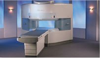
From Hitachi Medical Systems America, Inc.;
the AIRIS made its debut in 1995. Hitachi followed up with the AIRIS II system, which has proven equally successfully. 'All told, Hitachi has installed more than 1,000 MRI systems in the U.S., holding more than 17 percent of the total U.S. MRI installed base, and more than half of the installed base of open MR systems,' says Antonio Garcia, Frost and Sullivan industry research analyst.
Now Altaire employs a blend of innovative Hitachi features called VOSIā¢ technology, optimizing each sub-system's performance in concert with the
other sub-systems, to give the seamless mix of high-field performance
and the patient comfort, especially for claustrophobic patients, of open MR systems.
Device Information and Specification
CLINICAL APPLICATION
Whole body
DualQuad T/R Body Coil, MA Head, MA C-Spine, MA Shoulder, MA Wrist, MA CTL Spine, MA Knee, MA TMJ, MA Flex Body (3 sizes), Neck, small and large Extremity, PVA (WIP), Breast (WIP), Neurovascular (WIP), Cardiac (WIP) and MA Foot//Ankle (WIP)
SE, GE, GR, IR, FIR, STIR, ss-FSE, FSE, DE-FSE/FIR, FLAIR, ss/ms-EPI, ss/ms EPI- DWI, SSP, MTC, SE/GE-EPI, MRCP, SARGE, RSSG, TRSG, BASG, Angiography: CE, PC, 2D/3D TOF
IMAGING MODES
Single, multislice, volume study
TR
SE: 30 - 10,000msec GE: 3.6 - 10,000msec IR: 50 - 16,700msec FSE: 200 - 16,7000msec
TE
SE : 8 - 250msec IR: 5.2 -7,680msec GE: 1.8 - 2,000 msec FSE: 5.2 - 7,680
0.05 sec/image (256 x 256)
2D: 2 - 100 mm; 3D: 0.5 - 5 mm
Level Range: -2,000 to +4,000
COOLING SYSTEM TYPE
Water-cooled
3.1 m lateral, 3.6 m vertical
| |  | |
• View the DATABASE results for 'Altaire™' (2).
| | | | |  Further Reading: Further Reading: | News & More:
|
|
| |
|  |  | Searchterm 'MRI' was also found in the following services: | | | | |
|  |  |
| |
|
The brain tissue is provided with a tight endothelial layer on vessels that acts as a filter for substances that reach the brain through the blood stream. This filter is called the blood brain barrier.
The blood brain barrier is responsible for the absence of contrast agent enhancement in normal brain tissue after administration of the iodinated or paramagnetic contrast media used in brain MRI and computed tomography (CT) diagnostic. The absence of contrast uptake in normal tissue provides the basis for differentiation from pathological brain tissue, which is conversely characterized by a disruption of the blood brain barrier.
See also Contrast Enhanced MRI, MRI Safety, Adverse Reaction. | | | |  | |
• View the DATABASE results for 'Blood Brain Barrier' (7).
| | | | |  Further Reading: Further Reading: | News & More:
|
|
| |
|  | |  |  |
|  | | |
|
| |
 | Look
Ups |
| |