 | Info
Sheets |
| | | | | | | | | | | | | | | | | | | | | | | | |
 | Out-
side |
| | | | |
|
| | | | | |  | Searchterm 'Magnetic Resonance' was also found in the following services: | | | | |
|  |  |
| |
|

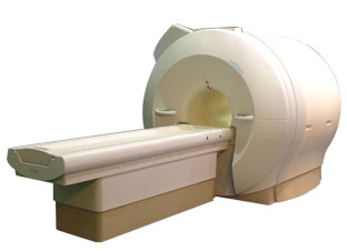
'Next generation MRI system 1.5T CHORUS developed by ISOL Technology is optimized for both clinical diagnostic imaging and for research development.
CHORUS offers the complete range of feature oriented advanced imaging techniques- for both clinical routine and research. The compact short bore magnet, the patient friendly design and the gradient technology make the innovation to new degree of perfection in magnetic resonance.'
Device Information and Specification
CLINICAL APPLICATION
Whole body
Spin Echo, Gradient Echo, Fast Spin Echo,
Inversion Recovery ( STIR, Fluid Attenuated Inversion Recovery), FLASH, FISP, PSIF, Turbo Flash ( MPRAGE ),TOF MR Angiography, Standard echo planar imaging package (SE-EPI, GE-EPI), Optional:
Advanced P.A. Imaging Package (up to 4 ch.), Advanced echo planar imaging package,
Single Shot and Diffusion Weighted EPI, IR/FLAIR EPI
STRENGTH
20 mT/m (Upto 27 mT/m)
| |  | | | |
|  |  | Searchterm 'Magnetic Resonance' was also found in the following services: | | | | |
|  |  |
| |
|
In the last years, cardiac MRI techniques have progressively improved. No other noninvasive imaging modality provides the same degree of contrast and temporal resolution for the assessment of cardiovascular anatomy and pathology. Contraindications MRI are the same as for other magnetic resonance techniques.
The primary advantage of MRI is extremely high contrast resolution between different tissue types, including blood. Moreover, MRI is a true 3 dimensional imaging modality and images can be obtained in any oblique plane along the true cardiac axes while preserving high temporal and spatial resolution with precise demonstration of cardiac anatomy without the administration of contrast media.
Due to these properties, MRI can precisely characterize cardiac function and quantify cavity volumes, ejection fraction, and left ventricular mass. In addition, cardiac MRI has the ability to quantify flow (see flow quantification), including bulk flow in vessels, pressure gradients across stenosis, regurgitant fractions and shunt fractions. Valve morphology and area can be determined and the severity of stenosis quantified. In certain disease states, such as myocardial infarction, the contrast resolution of MRI is further improved by the addition of extrinsic contrast agents (see myocardial late enhancement).
A dedicated cardiac coil, and a field strength higher than 1 Tesla is recommended to have sufficient signal. Cardiac MRI acquires ECG gating. Cardiac gating (ECGs) obtained within the MRI scanner, can be degraded by the superimposed electrical potential of flowing blood in the magnetic field. Therefore, excellent contact between the skin and ECG leads is necessary. For male patients, the skin at the lead sites can be shaved. A good cooperation of the patient is necessary because breath holding at the end of expiration is practiced during the most sequences.
See also Displacement Encoding with Stimulated Echoes.
For Ultrasound Imaging (USI) see Cardiac Ultrasound at Medical-Ultrasound-Imaging.com.
See also the related poll results: ' In 2010 your scanner will probably work with a field strength of' and ' MRI will have replaced 50% of x-ray exams by' | | | |  | |
• View the DATABASE results for 'Cardiac MRI' (15).
| | |
• View the NEWS results for 'Cardiac MRI' (15).
| | | | |  Further Reading: Further Reading: | | Basics:
|
|
News & More:
|  |
MRI technology visualizes heart metabolism in real time
Friday, 18 November 2022 by medicalxpress.com |  |  |
Even early forms of liver disease affect heart health, Cedars-Sinai study finds
Thursday, 8 December 2022 by www.eurekalert.org |  |  |
MRI sheds light on COVID vaccine-associated heart muscle injury
Tuesday, 15 February 2022 by www.sciencedaily.com |  |  |
Radiologists must master cardiac CT, MRI to keep pace with demand: The heart is not a magical organ
Monday, 1 March 2021 by www.radiologybusiness.com |  |  |
Diffusion weighted imaging (DWI) and diffusion tensor imaging (DTI) in the heart (myocardium)
Sunday, 30 August 2020 by github.com |  |  |
Non-invasive diagnostic procedures for suspected CHD: Search reveals informative evidence
Wednesday, 8 July 2020 by medicalxpress.co |  |  |
Cardiac MRI Becoming More Widely Available Thanks to AI and Reduced Exam Times
Wednesday, 19 February 2020 by www.dicardiology.com |  |  |
Controlling patient's breathing makes cardiac MRI more accurate
Friday, 13 May 2016 by www.upi.com |  |  |
Precise visualization of myocardial injury: World's first patient-based cardiac MRI study using 7T MRI
Wednesday, 10 February 2016 by medicalxpress.com |  |  |
New technique could allow for safer, more accurate heart scans
Thursday, 10 December 2015 by www.gizmag.com |
|
| |
|  | |  |  |  |
| |
|
A pacemaker is a device for internal or external battery-operated cardiac pacing to overcome cardiac arrhythmias or heart block. All implanted electronic devices are susceptible to the electromagnetic fields used in magnetic resonance imaging. Therefore, the main magnetic field, the gradient field, and the radio frequency (RF) field are potential hazards for cardiac pacemaker patients.
The pacemaker's susceptibility to static field and its critical role in life support have warranted special consideration. The static magnetic field applies force to magnetic materials. This force and torque effects rise linearly with the field strength of the MRI machines. Both, RF fields and pulsed gradients can induce voltages in circuits or on the pacing lead, which will heat up the tissue around e.g. the lead tip, with a potential risk of thermal injury.
Regulations for pacemakers provide that they have to switch to the magnet mode in static magnetic fields above 1.0 mT. In MR imaging, the gradient and RF fields may mimic signals from the heart with inhibition or fast pacing of the heart. In the magnet mode, most of the current pacemakers will pace with a fix pulse rate because they do not accept the heartsignals. However, the state of an implanted pacemaker will be unpredictable inside a strong magnetic field. Transcutaneous controller adjustment of pacing rate is a feature of many units. Some achieve this control using switches activated by the external application of a magnet to open/close the switch. Others use rotation of an external magnet to turn internal controls. The fringe field around the MRI magnet can activate such switches or controls. Such activations are a safety risk.
Areas with fields higher than 0.5 mT ( 5 Gauss Limit) commonly have restricted access and/or are posted as a safety risk to persons with pacemakers.

A Cardiac pacemaker is because the risks, under normal circumstances an absolute contraindication for MRI procedures.
Nevertheless, with special precaution the risks can be lowered. Reprogramming the pacemaker to an asynchronous mode with fix pacing rate or turning off will reduce the risk of fast pacing or inhibition. Reducing the SAR value reduces the potential MRI risks of heating. For MRI scans of the head and the lower extremities, tissue heating also seems to be a smaller problem. If a transmit receive coil is used to scan the head or the feet, the cardiac pacemaker is outside the sending coil and possible heating is very limited. | |  | |
• View the DATABASE results for 'Cardiac Pacemaker' (6).
| | | | |  Further Reading: Further Reading: | | Basics:
|
|
News & More:
| |
| |
|  |  | Searchterm 'Magnetic Resonance' was also found in the following services: | | | | |
|  |  |
| |
|
Quick Overview Please note that there are different common names for this artifact.
DESCRIPTION
Black or bright band
During frequency encoding, fat protons precess slower than water protons in the same slice because of their magnetic shielding. Through the difference in resonance frequency between water and fat, protons at the same location are misregistrated (dislocated) by the Fourier transformation, when converting MRI signals from frequency to spatial domain. This chemical shift misregistration cause accentuation of any fat-water interfaces along the frequency axis and may be mistaken for pathology. Where fat and water are in the same location, this artifact can be seen as a bright or dark band at the edge of the anatomy.
Protons in fat and water molecules are separated by a chemical shift of about 3.5 ppm. The actual shift in Hertz (Hz) depends on the magnetic field strength of the magnet being used. Higher field strength increases the misregistration, while in contrast a higher gradient strength has a positive effect. For a 0.3 T system operating at 12.8 MHz the shift will be 44.8 Hz compared with a 223.6 Hz shift for a 1.5 T system operating at 63.9 MHz.

Image Guidance
| |  | |
• View the DATABASE results for 'Chemical Shift Artifact' (7).
| | | | |  Further Reading: Further Reading: | Basics:
|
|
News & More:
| |
| |
|  |  | Searchterm 'Magnetic Resonance' was also found in the following services: | | | | |
|  |  |
| |
|
A large network of interconnecting blood vessels at the base of the brain that when visualized resembles a circle, the arteries effectively act as anastomoses for each other. This means that if any one of the communicating arteries becomes blocked, blood can flow from another part of the circle to ensure that blood flow is not compromised.
The circle of Willis is formed by both the internal carotid arteries, entering the brain from each side and the basilar artery, entering posteriorly. The connection of the vertebral arteries forms the basilar artery. The basilar artery divides into the right and left posterior cerebral arteries.
The internal carotid arteries trifurcate into the anterior cerebral artery, middle cerebral artery, and posterior communicating artery.
The two anterior cerebral arteries are joined together anteriorly by the anterior communicating artery. The posterior communicating arteries join the posterior cerebral arteries, completing the circle of Willis. The time of flight angiography MRI technique allows imaging of the circle of Willis without the need of a contrast medium (best results with high field MRI). A cerebrovasular contrast enhanced magnetic resonance angiography ( MRA) depicts the circle of Willis in addition to the vessels of the neck (carotid and vertebral arteries) with one bolus injection of a contrast agent.
For Ultrasound Imaging (USI) see Cerebrovascular Ultrasonography at Medical-Ultrasound-Imaging.com. | | | |  | |
• View the DATABASE results for 'Circle of Willis' (5).
| | | | |  Further Reading: Further Reading: | News & More:
|
|
| |
|  | |  |  |
|  | | |
|
| |
 | Look
Ups |
| |