 | Info
Sheets |
| | | | | | | | | | | | | | | | | | | | | | | | |
 | Out-
side |
| | | | |
|
| | | | |
Result : Searchterm 'Magnetic Shielding' found in 1 term [ ] and 10 definitions [ ] and 10 definitions [ ], (+ 8 Boolean[ ], (+ 8 Boolean[ ] results ] results
| previous 6 - 10 (of 19) nextResult Pages :  [1] [1]  [2 3] [2 3]  [4] [4] |  | |  | Searchterm 'Magnetic Shielding' was also found in the following services: | | | | |
|  |  |
| |
|

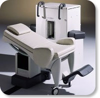
Developed by GE Lunar; the ARTOSCAN™-M is designed specifically for in-office musculoskeletal imaging. ARTOSCAN-M's compact, modular design allows placing within a clinical environment, bringing MRI to the patient. Patients remain outside the magnet at all times during the examinations, enabling constant patient-technologist contact. ARTOSCAN-M requires no special RF room, magnetic shielding, special power supply or air conditioning.
The C-SCAN™ (also known as Artoscan C) is developed from the ARTOSCAN™ - M, with a new computer platform.
Device Information and Specification
CLINICAL APPLICATION
Dedicated extremity
SE, GE, IR, STIR, FSE, 3D CE, GE-STIR, 3D GE, ME, TME, HSE
SLICE THICKNESS
2D: 2 mm - 10 mm;
3D: 0.6 mm - 10 mm
4,096 gray lvls, 256 lvls in 3D
POWER REQUIREMENTS
100/110/200/220/230/240V
| |  | | | | | | | | |
|  | |  |  |  |
| |
|
Quick Overview Please note that there are different common names for this artifact.
DESCRIPTION
Black or bright band
During frequency encoding, fat protons precess slower than water protons in the same slice because of their magnetic shielding. Through the difference in resonance frequency between water and fat, protons at the same location are misregistrated (dislocated) by the Fourier transformation, when converting MRI signals from frequency to spatial domain. This chemical shift misregistration cause accentuation of any fat-water interfaces along the frequency axis and may be mistaken for pathology. Where fat and water are in the same location, this artifact can be seen as a bright or dark band at the edge of the anatomy.
Protons in fat and water molecules are separated by a chemical shift of about 3.5 ppm. The actual shift in Hertz (Hz) depends on the magnetic field strength of the magnet being used. Higher field strength increases the misregistration, while in contrast a higher gradient strength has a positive effect. For a 0.3 T system operating at 12.8 MHz the shift will be 44.8 Hz compared with a 223.6 Hz shift for a 1.5 T system operating at 63.9 MHz.

Image Guidance
| |  | |
• View the DATABASE results for 'Chemical Shift Artifact' (7).
| | | | |  Further Reading: Further Reading: | | Basics:
|
|
News & More:
| |
| |
|  | |  |  |  |
| |
|


O-scan is manufactured and distributed by Esaote SpA
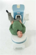
O-scan is a compact, dedicated extremity MRI system designed for easy installation and high throughput. The complete system fits in a 9' x 10' room, doesn't need for RF or magnetic shielding and it plugs in the wall. The 0.31T permanent magnet along with dual phased array RF coils, and advanced imaging protocols provide outstanding image quality and fast 25 minute complete examinations.
Esaote North America is the exclusive distributor of the O-scan system in the USA.
Device Information and Specification CLINICAL APPLICATION Dedicated Extremity
PULSE SEQUENCES
SE, HSE, HFE, GE, 2dGE, ME, IR, STIR, Stir T2, GESTIR, TSE, TME, FSE STIR, FSE ( T1, T2), X-Bone, Turbo 3DT1, 3D SHARC, 3D SST1, 3D SST2 2D: 2mm - 10 mm, 3D: 0.6 - 10 mm POWER REQUIREMENTS 100/110/200/220/230/240 | |  | | | |
|  |  | Searchterm 'Magnetic Shielding' was also found in the following services: | | | | |
|  |  |
| |
|
Radio frequency shielding includes the construction of enclosures for the purpose of reducing the transmission of electric or magnetic fields from one space to another ( Faraday cage, Faraday shield). Electrically conducted shielding is designed to isolate MRI systems from its environment at the resonant frequencies.
All electronic and computer systems radiate certain frequencies of radio and magnetic waves. They can interfere with other equipment in the vicinity. Magnetic shielding enclosures are used to reduce the levels of RF radiation that enters or leaves the shielded room.
Copper shielding enclosures are designed to filter a range of frequencies under specified conditions. One of the characteristics of copper is its high electrical conductivity. Also its other physical properties like ductility, malleability, and ease of soldering, make it an ideal material for radio frequency shielding. Sheet copper can be formed into any shape and size, and electrically connected to a grounding system to provide an effective RF shielding.
See also MRI Safety | |  | |
• View the DATABASE results for 'Radio Frequency Shielding' (3).
| | | | |
|  | |  |  |  |
| |
|
| |  | |
• View the DATABASE results for 'Shielding' (27).
| | | | |  Further Reading: Further Reading: | News & More:
|
|
| |
|  | |  |  |
|  | |
|  | | |
|
| |
 | Look
Ups |
| |