Magnetic Resonance - Technology Information Portal Welcome to MRI Technology
Info
  Sheets

Out-
      side


 'Proton' 
SEARCH FOR    
 
  2 3 5 A B C D E F G H I J K L M N O P Q R S T U V W X Y Z
Result : Searchterm 'Proton' found in 3 terms [] and 82 definitions []
previous     71 - 75 (of 85)     next
Result Pages : [1]  [2 3 4 5 6 7 8 9 10 11 12 13 14 15 16 17]
Searchterm 'Proton' was also found in the following services: 
spacer
News  (6)  Resources  (7)  Forum  (16)  
 
Saturation Recovery
 
(SR) Particular type of partial saturation pulse sequence in which the preceding pulses leave the spins in a state of saturation, so that recovery at the time of the next pulse has taken place from an initial condition of no magnetization. A rare used MRI pulse sequence that generates a predominantly proton density dependent signal, basically employing a 90° RF excitation pulse, with a very long repetition time. With this technique T1 times can be measured faster than with inversion recovery pulse sequences.
This saturation recovery sequence consists of multiple 90° radio frequency (RF) pulses with a short repetition time. A spoiler gradient pulse dephases the longitudinal magnetization that remains after the first 90° radio frequency pulse. A repetition time interval after the application of this spoiling gradient turns an additional 90° pulse the new developed longitudinal magnetization into the transverse plane, followed by recording a gradient echo.
spacer
 
• Related Searches:
    • Fat Saturation
    • Spectral Presaturation Inversion Recovery
    • Saturation
    • Suppression
    • Gradient Echo Sequence
 
Further Reading:
  Basics:
Contrast mechanisms in magnetic resonance imaging
2004   by www.iop.org    
Searchterm 'Proton' was also found in the following service: 
spacer
Radiology  (32) Open this link in a new window
Short T1-Relaxation Gastrointestinal AgentsInfoSheet: - Contrast Agents - 
Intro, Overview, 
Characteristics, 
Types of, 
etc.
 
Protons in -CH2- groups, e.g., contained in fatty emulsions, mineral or vegetable oil or sucrose polyester, have a fast relaxation and short T1 time. These agents with short T1-relaxation, if used in gastrointestinal imaging, produce bright signal intensities in the intestine on T1 weighted sequences. Palatable oil emulsions can produce appropriate contrast opacification of the stomach as well as the small bowel, but caused by absorption in the distal small bowel these materials are not suitable for use in MRI colonoscopy.

See also Positive Oral Contrast Agents and Gastrointestinal Paramagnetic Contrast Agents.
 
Images, Movies, Sliders:
 MR Colonography Gadolinium per Rectum  Open this link in a new window
      

Courtesy of  Robert R. Edelman
 
spacer

• View the DATABASE results for 'Short T1-Relaxation Gastrointestinal Agents' (2).Open this link in a new window

MRI Resources 
Shoulder MRI - Diffusion Weighted Imaging - Guidance - MRI Technician and Technologist Career - Corporations - Homepages
 
Shoulder MRI
 
MRI of the shoulder with its excellent soft tissue discrimination, and high spatial resolution offers the best noninvasive way to study the shoulder. MRI images of the bone, muscles and tendons of the glenohumeral joint can be obtained in any oblique planes and projections. MRI gives excellent depiction of rotator cuff tears, injuries to the biceps tendon and damage to the glenoid labrum. Shoulder MRI is better than ultrasound imaging at depicting structural changes such as osteophytic spurs, ligament thickening, and acromial shape that may have predisposed to tendon degeneration.
A dedicated shoulder coil and careful patient positioning in external rotation with the shoulder as close as reasonably possible to the center of the magnet is necessary for a good image quality. If possible, the opposite shoulder should be lifted up, so that the patient lies on the imaged shoulder in order to rotate and fix this shoulder to reduce motion during breathing.
Axial, coronal oblique, and sagittal oblique proton density with fat suppression, T2 and T1 provide an assessment of the rotator cuff, biceps, deltoid, acromio-clavicular joint, the glenohumeral joint and surrounding large structures. If a labral injury is suspected, a Fat Sat gradient echo sequence is recommended. In some cases, a direct MR shoulder arthrogram with intra-articular injection of dilute gadolinium or an indirect arthrogram with imaging 20 min. after intravenous injection may be helpful.

See also Imaging of the Extremities.
 
Images, Movies, Sliders:
 Anatomic Imaging of the Shoulder  Open this link in a new window
      

Courtesy of  Robert R. Edelman

 
Radiology-tip.comradArthrography
spacer
Medical-Ultrasound-Imaging.comLow Intensity Pulsed Ultrasound,  Musculoskeletal and Joint Ultrasound
spacer

• View the DATABASE results for 'Shoulder MRI' (3).Open this link in a new window


• View the NEWS results for 'Shoulder MRI' (1).Open this link in a new window.
 
Further Reading:
  News & More:
The Spectrum of Shoulder Pathologies on Magnetic Resonance Imaging: A Pictorial Review
Wednesday, 6 September 2023   by www.cureus.com    
MRI costs wide-ranging
Thursday, 14 April 2011   by www.chieftain.com    
MRE Could Provide A Definitive Diagnosis For People With Muscle Pain, Study Shows
Friday, 30 November 2007   by www.sciencedaily.com    
Peer-Reviewed Study Concludes The FONAR UPRIGHT™ MRI Could Serve as the “Standard Procedure of Care” for Pediatric Shoulder Malady
Wednesday, 30 May 2007   by www.fonar.com    
Searchterm 'Proton' was also found in the following services: 
spacer
News  (6)  Resources  (7)  Forum  (16)  
 
Signa 3.0T™InfoSheet: - Devices -
Intro, 
Types of Magnets, 
Overview, 
etc.MRI Resource Directory:
 - Devices -
 
www.gehealthcare.com/usen/mr/s_excite3/index.html (Signa VH/i 3.0T)
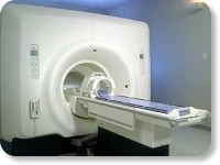
With GE Healthcare leading-edge technology in ultra-high-field imaging. The 3 T VH/i provides a platform for advanced applications in radiology, cardiology, psychology and psychiatry. Real-time image processing lets you acquire multislice whole brain images and map brain functions for research or surgical planning. And the 3 T Signa VH/i is flexible enough to provide clinicians with high performance they require. It can provide not only outstanding features in brain scanning and neuro-system research, but also a wide range of use in scanning breasts, extremities, the spine and the cardiovascular systems.
Device Information and Specification
CLINICAL APPLICATION
Whole body
CONFIGURATION
Cylindrical - high homogeneity
T/R quadrature head, T/R quadrature body, T/R phased array extremity (opt)
SYNCHRONIZATION
ECG/peripheral, respiratory gating
PULSE SEQUENCES
SE, IR, 2D/3D GRE, FGRE, RF-spoiled GRE, FSE, Angiography: 2D/3D TOF, 2D/3D phase contrast vascular
IMAGING MODES
Single, multislice, volume study, fast scan, multi slab, cine, localizer
SINGLE SLICE
100 Images/sec with Reflex100
MULTISLICE
100 Images/sec with Reflex100
1 cm to 40 cm continuous
2D 0.5-100mm in 0.1mm incremental
1280 x 1024
MEASURING MATRIX
128x512 steps 32 phase encode
PIXEL INTENSITY
256 gray levels
55cm
MAGNET WEIGHT
15102 kg incl. cryogen's
H*W*D
260cm x 238cm x 265cm
POWER REQUIREMENTS
480 or 380/415, 3 phase ||
COOLING SYSTEM TYPE
Closed-loop water-cooled grad.
Less than 0.14 L/hr liquid He
STRENGTH
40mT/m
5-GAUSS FRINGE FIELD, radial/axial
5.4 m x 3.2 m
Superconductive + hi order active
spacer

• View the DATABASE results for 'Signa 3.0T™' (2).Open this link in a new window

Searchterm 'Proton' was also found in the following service: 
spacer
Radiology  (32) Open this link in a new window
Signa HDx 3.0T™InfoSheet: - Devices -
Intro, 
Types of Magnets, 
Overview, 
etc.
 
gehealthcare.com/euen/mri/products/signa-hdx-3t/index.html From GE Healthcare;
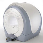
The Signa HDx MRI system is GE's leading edge whole body magnetic resonance scanner designed to support high resolution, high signal to noise ratio, and short scan times.
Signa HDx 3.0T offers new technologies like ultra-fast image reconstruction through the new XVRE recon engine, advancements in parallel imaging algorithms and the broadest range of premium applications. The HD applications, PROPELLER (high-quality brain imaging extremely resistant to motion artifacts), TRICKS (contrast-enhanced angiographic vascular lower leg imaging), VIBRANT (for breast MRI), LAVA (high resolution liver imaging with shorter breath holds and better organ coverage) and MR Echo (high-definition cardiac images in real time) offer unique capabilities.
Device Information and Specification
CLINICAL APPLICATION
Whole body
CONFIGURATION
Compact short bore
Head and body coil, T/R quadrature head; optional coils e.g., T/R phased array extremity abdomen, spine, breast, knee, shoulder, cardiac imaging coils
SYNCHRONIZATION
ECG/peripheral, respiratory gating
PULSE SEQUENCES
SE, IR, 2D/3D GRE, RF-spoiled GRE, 2DFGRE, 2DFSPGR, 3DFGRE, 3DFSPGR, 3DTOFGRE, 3DFSPGR, 2DFSE, 2DFSE-XL, 2DFSE-IR, T1-FLAIR, SSFSE, EPI, DW-EPI, BRAVO, Angiography: 2D/3D TOF, 2D/3D phase contrast vascular
IMAGING MODES
Single, multislice, volume study, fast scan, multi slab, cine, localizer
1 cm to 40 cm continuous
2D 0.5 mm; 3D 0.1 mm
1024 x 1024
PIXEL INTENSITY
256 gray levels
60 cm
MAGNET WEIGHT
12000 kg
H*W*D
240 x 2216,6 x 201,6 cm
POWER REQUIREMENTS
480 or 380/415, 3 phase ||
COOLING SYSTEM TYPE
Closed-loop water-cooled grad.
0.03 L/hr helium
STRENGTH
23 - 50 mT/m
80 - 150 mT/m/ms
5-GAUSS FRINGE FIELD
2.8 m / 5.0 m
second and high order
spacer
MRI Resources 
Lung Imaging - Contrast Agents - Absorption and Emission - Abdominal Imaging - Knee MRI - Veterinary MRI
 
previous      71 - 75 (of 85)     next
Result Pages : [1]  [2 3 4 5 6 7 8 9 10 11 12 13 14 15 16 17]
 Random Page
 
Share This Page
FacebookTwitterLinkedIn

MR-TIP    
Community   
User
Pass
Forgot your UserID/Password ?    


Next big thing in MRI will be :
AI 
remote operator 
personalized protocols 
helium-free 
molecular MRI 
portable MRI 

Look
      Ups


MR-TIP.com uses cookies! By browsing MR-TIP.com, you agree to our use of cookies.

Magnetic Resonance - Technology Information Portal
Member of SoftWays' Medical Imaging Group - MR-TIP • Radiology-TIP • Medical-Ultrasound-Imaging • 
Copyright © 2003 - 2024 SoftWays. All rights reserved. [ 3 December 2024]
Terms of Use | Privacy Policy | Advertising
 [last update: 2024-02-26 03:41:00]