Magnetic Resonance - Technology Information Portal Welcome to MRI Technology
Info
  Sheets

Out-
      side


 'Pulse, 180' 
SEARCH FOR    
 
  2 3 5 A B C D E F G H I J K L M N O P Q R S T U V W X Y Z
Result : Searchterm 'Pulse, 180' found in 1 term [] and 0 definition [], (+ 10 Boolean[] results
1 - 5 (of 11)     next
Result Pages : [1]  [2 3]
MRI Resources 
Coils - Diffusion Weighted Imaging - Shielding - MRCP - Liver Imaging - MRI Technician and Technologist Schools
 
Pulse, 180°
 
RF pulse designed to rotate the macroscopic magnetization vector 180° in space as referred to the rotating frame of reference, usually about an axis at right angles to the main magnetic field. If the spins are initially aligned with the magnetic field, this pulse will produce inversion. Also called P Pulse.
spacer
 
• Share the entry 'Pulse, 180°':  Facebook  Twitter  LinkedIn  
 
Further Reading:
  Basics:
Clinical evaluation of a speed optimized T2 weighted fast spin echo sequence at 3.0 T using variable flip angle refocusing, half-Fourier acquisition and parallel imaging
Wednesday, 25 October 2006
MRI Resources 
Colonography - Breast Implant - Online Books - Stimulator pool - Anatomy - Quality Advice
 
Ultrafast Gradient Echo SequenceInfoSheet: - Sequences - 
Intro, 
Overview, 
Types of, 
etc.MRI Resource Directory:
 - Sequences -
 
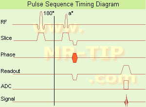
Ultrafast Gradient Echo Sequence Timing Diagram In simple ultrafast GRE imaging, TR and TE are so short, that tissues have a poor imaging signal and - more importantly - poor contrast except when contrast media enhanced (contrast enhanced angiography). Therefore, the magnetization is 'prepared' during the preparation module, most frequently by an initial 180° inversion pulse.
In the pulse sequence timing diagram, the basic ultrafast gradient echo sequence is illustrated. The 180° inversion pulse is executed one time (to the left of the vertical line), the right side represents the data collection period and is often repeated depending on the acquisition parameters.
See also Pulse Sequence Timing Diagram, there you will find a description of the components.
Ultrafast GRE sequences have a short TR,TE, a low flip angle and TR is so short that image acquisition lasts less than 1 second and typically less than 500 ms. Common TR: 3-5 msec, TE: 2 msec, and the flip angle is about 5°. Such sequences are often labeled with the prefix 'Turbo' like TurboFLASH, TurboFFE and TurboGRASS.
This allows one to center the subsequent ultrafast GRE data acquisition around the inversion time TI, where one of the tissues of interest has very little signal as its z-magnetization is passing through zero.
Unlike a standard inversion recovery (IR) sequence, all lines or a substantial segment of k-space image lines are acquired after a single inversion pulse, which can then together be considered as readout module. The readout module may use a variable flip angle approach, or the data acquisition may be divided into multiple segments (shots). The latter is useful particularly in cardiac imaging where acquiring all lines in a single segment may take too long relative to the cardiac cycle to provide adequate temporal resolution.
If multiple lines are acquired after a single pulse, the pulse sequence is a type of gradient echo echo planar imaging (EPI) pulse sequence.

See also Magnetization Prepared Rapid Gradient Echo (MPRAGE) and Turbo Field Echo (TFE).
spacer

• View the DATABASE results for 'Ultrafast Gradient Echo Sequence' (13).Open this link in a new window

MRI Resources 
MRCP - MRI Technician and Technologist Career - Claustrophobia - Equipment - Databases - Knee MRI
 
Image Selected In Vivo Spectroscopy
 
(ISIS) Image selected in vivo spectroscopy is used as a localization sequence to provide complete gradient controlled three-dimensional localization with a reduced number of sequence cycles, e.g. for in vivo 31P spectroscopy. The ISIS method generates three 180° pulses prior to a 90° pulse, after which the free induction decay is recorded. Specific 180° pulses (slice-selective) are combined and the FID's added or subtracted to generate a spectrum.
An advantage of the ISIS method is that the magnetization (before the final 90° pulse) is predominantly along the z-axis and so T2 effects are relatively small. This explains the value of this technique for 31P data acquisition, because some phosphorus metabolites (e.g. ATP) have short T2 values.
A disadvantage is that eight acquisitions are required to accomplish the spatial localization, therefore the sequence cannot be used for localized shimming. Another problem, because any variation between these data collections (for example, due to movement) will degrade these applications, can be solved by incorporating outer volume suppression techniques such as OSIRIS (modified ISIS).
spacer
MRI Resources 
Stent - DICOM - Manufacturers - Hospitals - Implant and Prosthesis - Bioinformatics
 
Spin EchoForum -
related threads
 
(SE) The Reappearance of the MR signal after the FID has apparently died away, as a result of the effective reversal (rephasing) of the dephasing spins by techniques such as specific RF pulse sequences or pairs of field gradient pulses, applied in time shorter than or on the order of T2. Proper selection of the TE time of the pulse sequence can help to control the amount of T1 or T2 contrast present in the image. Pulse sequences of the spin echo type, usually employs a 90° pulse, followed by one or more 180° pulses to eliminate field inhomogeneity and chemical shift effects at the echo. Caused by this 180° refocusing pulse, spin echo or fast spin echo (FSE, TSE) sequences are more robust against e.g. susceptibility artifacts than sequences of the gradient echo type.
spacer

• View the DATABASE results for 'Spin Echo' (96).Open this link in a new window


• View the NEWS results for 'Spin Echo' (1).Open this link in a new window.
 
Further Reading:
  Basics:
Spin echoes, CPMG and T2 relaxation - Introductory NMR & MRI from Magritek
2013   by www.azom.com    
  News & More:
EVALUATION OF HUMAN STROKE BY MR IMAGING
2000
MRI Resources 
RIS - Lung Imaging - MRI Accidents - MRI Technician and Technologist Schools - Brain MRI - Chemistry
 
Spin Echo Multi SliceInfoSheet: - Sequences - 
Intro, 
Overview, 
Types of, 
etc.MRI Resource Directory:
 - Sequences -
 
(SEMS) This pulse sequence is composed of a 90° RF pulse followed by a 180° refocusing pulse. Both RF pulses are applied in the presence of a slice select gradient.
By choosing of different TR and TE, depending on the T1 and T2 values of the tissues, proton density, T1 weighted and T2 weighted images can be acquired.
The inversion recovery option enlarge the RF pulses with a 180° inverting pulse, applied a TI time before the beginning of the pulse sequence in order to manipulate image contrast.
See also Spin Echo Sequence.
spacer
 
Further Reading:
  Basics:
Fast Spin Echo(.pdf)
Tuesday, 24 January 2006   by www.81bones.net    
MRI Resources 
Abdominal Imaging - Societies - - Spectroscopy pool - MR Myelography - MRA
 
     1 - 5 (of 11)     next
Result Pages : [1]  [2 3]
 Random Page
 
Share This Page
FacebookTwitterLinkedIn

MR-TIP    
Community   
User
Pass
Forgot your UserID/Password ?    


New acceleration techniques will :
reduce scan times 
cause artifacts 
increase expenses 
be useful if you have a lot of experience 
doesn't do much 
never heard of 

Look
      Ups


MR-TIP.com uses cookies! By browsing MR-TIP.com, you agree to our use of cookies.

Magnetic Resonance - Technology Information Portal
Member of SoftWays' Medical Imaging Group - MR-TIP • Radiology-TIP • Medical-Ultrasound-Imaging • 
Copyright © 2003 - 2024 SoftWays. All rights reserved. [ 21 November 2024]
Terms of Use | Privacy Policy | Advertising
 [last update: 2024-02-26 03:41:00]