 | Info
Sheets |
| | | | | | | | | | | | | | | | | | | | | | | | |
 | Out-
side |
| | | | |
|
| | | | |
Result : Searchterm 'Resistive Magnet' found in 1 term [ ] and 2 definitions [ ] and 2 definitions [ ], (+ 2 Boolean[ ], (+ 2 Boolean[ ] results ] results
| 1 - 5 (of 5) Result Pages :  [1] [1] |  | | |  |  |  |
| |
|
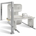
A type of magnet that utilizes the principles of electromagnetism to generate the magnetic field. Typically large current values and significant cooling of the magnet coils is required. The resistive magnet does not require cryogens, but needs a constant power supply to maintain a homogenous magnetic field, and can be quite expensive to maintain.
Resistive magnets fall into two general categories - iron-core and air-core.
Iron-core electromagnets provide the advantages of a vertically oriented magnetic field, and a limited fringe field with little, if any, missile effects due to the closed iron-flux return path.
Air-core electromagnets exhibit horizontally oriented fields, which have large fringe fields (unless magnetically shielded) and are prone to missile effects. Resistive magnets are typically limited to maximum field strengths of approximately 0.6T. | |  | | | | • Share the entry 'Resistive Magnet':    | | | | | | | | | |
|  | |  |  |  |
| |
|
Magnetic resonance imaging ( MRI) is based on the magnetic resonance phenomenon, and is used for medical diagnostic imaging since ca. 1977 (see also MRI History).
The first developed MRI devices were constructed as long narrow tunnels. In the meantime the magnets became shorter and wider. In addition to this short bore magnet design, open MRI machines were created. MRI machines with open design have commonly either horizontal or vertical opposite installed magnets and obtain more space and air around the patient during the MRI test.
The basic hardware components of all MRI systems are the magnet, producing a stable and very intense magnetic field, the gradient coils, creating a variable field and radio frequency (RF) coils which are used to transmit energy and to encode spatial positioning. A computer controls the MRI scanning operation and processes the information.
The range of used field strengths for medical imaging is from 0.15 to 3 T. The open MRI magnets have usually field strength in the range 0.2 Tesla to 0.35 Tesla. The higher field MRI devices are commonly solenoid with short bore superconducting magnets, which provide homogeneous fields of high stability.
There are this different types of magnets:
The majority of superconductive magnets are based on niobium-titanium (NbTi) alloys, which are very reliable and require extremely uniform fields and extreme stability over time, but require a liquid helium cryogenic system to keep the conductors at approximately 4.2 Kelvin (-268.8° Celsius). To maintain this temperature the magnet is enclosed and cooled by a cryogen containing liquid helium (sometimes also nitrogen).
The gradient coils are required to produce a linear variation in field along one direction, and to have high efficiency, low inductance and low resistance, in order to minimize the current requirements and heat deposition. A Maxwell coil usually produces linear variation in field along the z-axis; in the other two axes it is best done using a saddle coil, such as the Golay coil.
The radio frequency coils used to excite the nuclei fall into two main categories; surface coils and volume coils.
The essential element for spatial encoding, the gradient coil sub-system of the MRI scanner is responsible for the encoding of specialized contrast such as flow information, diffusion information, and modulation of magnetization for spatial tagging.
An analog to digital converter turns the nuclear magnetic resonance signal to a digital signal. The digital signal is then sent to an image processor for Fourier transformation and the image of the MRI scan is displayed on a monitor.
For Ultrasound Imaging (USI) see Ultrasound Machine at Medical-Ultrasound-Imaging.com.
See also the related poll results: ' In 2010 your scanner will probably work with a field strength of' and ' Most outages of your scanning system are caused by failure of' | | | | | | | | |
• View the DATABASE results for 'Device' (141).
| | |
• View the NEWS results for 'Device' (29).
| | | | |  Further Reading: Further Reading: | News & More:
|
 |
small-steps-can-yield-big-energy-savings-and-cut-emissions-mris
Thursday, 27 April 2023 by www.itnonline.com |  |  |
Portable MRI can detect brain abnormalities at bedside
Tuesday, 8 September 2020 by news.yale.edu |  |  |
Point-of-Care MRI Secures FDA 510(k) Clearance
Thursday, 30 April 2020 by www.diagnosticimaging.com |  |  |
World's First Portable MRI Cleared by FDA
Monday, 17 February 2020 by www.medgadget.com |  |  |
Low Power MRI Helps Image Lungs, Brings Costs Down
Thursday, 10 October 2019 by www.medgadget.com |  |  |
Cheap, portable scanners could transform brain imaging. But how will scientists deliver the data?
Tuesday, 16 April 2019 by www.sciencemag.org |  |  |
The world's strongest MRI machines are pushing human imaging to new limits
Wednesday, 31 October 2018 by www.nature.com |  |  |
Kyoto University and Canon reduce cost of MRI scanner to one tenth
Monday, 11 January 2016 by www.electronicsweekly.com |  |  |
A transportable MRI machine to speed up the diagnosis and treatment of stroke patients
Wednesday, 22 April 2015 by medicalxpress.com |  |  |
Portable 'battlefield MRI' comes out of the lab
Thursday, 30 April 2015 by physicsworld.com |  |  |
Chemists develop MRI technique for peeking inside battery-like devices
Friday, 1 August 2014 by www.eurekalert.org |  |  |
New devices doubles down to detect and map brain signals
Monday, 23 July 2012 by scienceblog.com |
|
| |
|  | |  |  |  |
| |
|
| |  | |
• View the DATABASE results for 'Electromagnet' (24).
| | |
• View the NEWS results for 'Electromagnet' (8).
| | | | |  Further Reading: Further Reading: | | Basics:
|
|
News & More:
| |
| |
|  | |  |  |  |
| |
|

From Philips Medical Systems;
the Panorama 0.23 T, providing a new design optimized for patient comfort, faster reconstruction time than before (300 images/second) and new gradient
specifications. Philips' Panorama 0.23 T I/T supports MR-guided interventions, resulting in minimally invasive procedures, more targeted surgery, reduced recovery time and shorter hospital stays. Optional OptoGuide functionality enables real-time needle tracking. Philips' Panorama 0.23 TPanorama 0.2 R/T is the first and only open MRI system to enable radiation therapy planning using MR data sets. The Panorama also features the new and consistent Philips User Interface, an essential element of the Vequion clinical IT family of products and services.
Device Information and Specification CLINICAL APPLICATION Whole body SE, FE, IR, FFE, DEFFE, DESE, TSE, DETSE, Single shot SE, DRIVE, Balanced FFE, MRCP, Fluid Attenuated Inversion Recovery, Turbo FLAIR, IR-TSE, T1-STIR TSE, T2-STIR TSE, Diffusion Imaging, 3D SE, 3D FFE, MTC;; Angiography: CE-ANGIO, MRA 2D, 3D TOFOpen x 46 cm x infinite (side-first patient entry) POWER REQUIREMENTS 400/480 V COOLING SYSTEM TYPE Closed loop chilled water ( chiller included) | |  | |
• View the DATABASE results for 'Panorama 0.23T™' (2).
| | | | |  Further Reading: Further Reading: | News & More:
|
|
| |
|  | |  |  |  |
| |
|
A quench is the rapid helium evaporation and the loss of superconductivity of the current-carrying coil that may occur unexpectedly, or from pressing the emergency button in a superconducting magnet. As the superconductive magnet becomes resistive, heat will be released that can result in boiling of liquid helium in the cryostat. This may present a hazard if not properly planned for.
The evaporated coolant requires emergency venting systems to protect patients and operators. Quenching can cause total magnet failure and cannot be stopped. MRI systems are designed such that all of the escaping cryogenic gas is directed out of the building ( quench pipe through the roof or the wall). In the event of a burst of the tank (possible in the case of an accident) or a blockage of the pipes, the helium gas will be forced into the scanner room, giving rise to a large white cloud of chilled gas. Under such circumstances it is essential that the scanner room is evacuated, also caused by the displacement of oxygen, which under extreme conditions could lead to asphyxiation. The force of quenching can be strong enough to destroy the walls of the scanner room or the MRI equipment. | |  | |
• View the DATABASE results for 'Quenching' (5).
| | | | |
|  | |  |  |
|  | 1 - 5 (of 5) Result Pages :  [1] [1] |
| |
|
| |
 | Look
Ups |
| |