 | Info
Sheets |
| | | | | | | | | | | | | | | | | | | | | | | | |
 | Out-
side |
| | | | |
|
| | | | | |  | Searchterm 'Resolution' was also found in the following services: | | | | |
|  |  |
| |
|
The partial volume effect is the loss of contrast between two adjacent tissues in an image caused by insufficient resolution so that more than one tissue type occupies the same voxel (or pixel). That may induce a partial volume artifact, dependent on the size of the image voxel. If fat and water spins occupy the same voxel, their signals interfere destructively. A small amount of water signal may be eliminated by a larger lipid signal from the same voxel, resulting in a voxel that appears to contain only lipid. The partial volume effect is minimal with thin slice thickness and sufficiently high resolution, so that fat and water or other different structures are unlikely to occupy the same voxel.

Image Guidance
| |  | | | |
|  |  | Searchterm 'Resolution' was also found in the following services: | | | | |
|  |  |
| |
|
| |  | |
• View the DATABASE results for 'Phased Array Coil' (9).
| | | | |  Further Reading: Further Reading: | Basics:
|
|
| |
|  | |  |  |  |
| |
|
(3D MRA) The 3D angiography technique can be applied to focus on fast flowing (arterial) blood and to visualize small tortuous vessels. 3D TOF images are less sensitive to turbulent flow artifacts.
The advantage of this approach is that the signal, acquired from the entire
volume has an increased signal to noise ratio. Slices are defined by a second phase encoded axis, which divides the volume into 'partitions'.
3D TOF MRA is acquired with 3D FT slabs or multiple overlapping thin 3D FT slabs ( MOTSA) depending on the coverage required and the range of flow-velocities under examination.
Such 3D techniques can provide equal spatial resolution along all three axes, i.e. be 'isotropic', or the partition thickness can be greater or less than the in plane spatial resolution in which case can be said to be 'anisotropic'.
The circle of Willis, anatomy as well as its fast arterial flow, lends itself well to both 3D TOF and 2D or 3D phase contrast angiography. | | | |  | |
• View the DATABASE results for '3 Dimensional Magnetic Resonance Angiography' (2).
| | | | |  Further Reading: Further Reading: | Basics:
|
|
| |
|  |  | Searchterm 'Resolution' was also found in the following services: | | | | |
|  |  |
| |
|
An array coil combines the advantages of smaller coils (high SNR) with those of larger coils (large measurement field).
This type of RF coil is composed of separate multiple smaller coils, which can be used individually ( switchable coil) or combined.
When used simultaneously, the elements can either be:
•
coupled array coils - electrically coupled to each other through common transmission lines or mutual inductance
•
isolated array coils - electrically isolated from each other with separate transmission lines and receivers and minimum effective
mutual inductance, and with the signals from each transmission line processed independently or at different frequencies
•
phased array coils - multiple small coils arranged to efficiently cover a specific anatomic region and obtain high-resolution, high-SNR images of a larger volume. The data from the individual coils is integrated by special software to produce the high-resolution images.
See also the related poll result: ' 3rd party coils are better than the original manufacturer coils'
| | | |  | |
• View the DATABASE results for 'Array Coil' (22).
| | |
• View the NEWS results for 'Array Coil' (1).
| | | | |  Further Reading: Further Reading: | News & More:
|
|
| |
|  |  | Searchterm 'Resolution' was also found in the following services: | | | | |
|  |  |
| |
|

From Aurora Imaging Technology, Inc.;
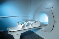
The Aurora® 1.5T Dedicated Breast MRI System with Bilateral SpiralRODEO™ is the first and only FDA approved MRI device designed specifically for breast imaging. The Aurora System, which is already in clinical use at a growing number of leading breast care centers in the US, Europe, got in December 2006 also the approval from the State Food and Drug Administration of the People's Republic of China (SFDA).
'Some of the proprietary and distinguishing features of the Aurora System include: 1) an ellipsoid magnetic shim that provides coverage of both breasts, the chest wall and bilateral axillary lymph nodes; 2) a precision gradient coil with the high linearity required for high resolution spiral reconstruction;; 3) a patient-handling table that provides patient comfort and procedural utility; 4) a fully integrated Interventional System for MRI guided biopsy and localization; and 5) the user-friendly AuroraCAD™ computer-aided image display system designed to improve the accuracy and efficiency of diagnostic interpretations.'
Device Information and Specification
CONFIGURATION
Short bore compact
TE
From 5 ms for RODEO Plus to over 80 ms, 120 ms for T2 sequences
Around 0.02 sec for a 256x256 image, 12.4 sec for a 512 x 512 x 32 multislice set
20 - 36 cm, max. elliptical 36 x 44 cm
POWER REQUIREMENTS
150A/120V-208Y/3 Phase//60 Hz/5 Wire
| |  | |
• View the DATABASE results for 'Aurora® 1.5T Dedicated Breast MRI System' (2).
| | |
• View the NEWS results for 'Aurora® 1.5T Dedicated Breast MRI System' (3).
| | | | |  Further Reading: Further Reading: | News & More:
|
|
| |
|  | |  |  |
|  | |
|  | | |
|
| |
 | Look
Ups |
| |