 | Info
Sheets |
| | | | | | | | | | | | | | | | | | | | | | | | |
 | Out-
side |
| | | | |
|
| | | | |
Result : Searchterm 'Saturation' found in 12 terms [ ] and 41 definitions [ ] and 41 definitions [ ] ]
| previous 21 - 25 (of 53) nextResult Pages :  [1 2 3] [1 2 3]  [4 5 6 7 8 9 10 11] [4 5 6 7 8 9 10 11] |  | |  | Searchterm 'Saturation' was also found in the following services: | | | | |
|  |  |
| |
|
Quick Overview
DESCRIPTION
Loss of signal of flowing blood

Image Guidance
| |  | | | | | | | | |  Further Reading: Further Reading: | Basics:
|
|
| |
|  |  | Searchterm 'Saturation' was also found in the following service: | | | | |
|  |  |
| |
|
(2D TOF MRA) This form of MR angiography is based on the acquisition of multiple, short-TR, gradient echo single slice images. 2D TOF MRA is the preferred technique for visualizing slow flow, how for example it happens in veins. 2D TOF MRA consists of multiple sequentially-acquired single slices, therefore the saturation effects are minimized. | |  | | | |
|  | |  |  |  |
| |
|
Quick Overview
Please note that there are different common names for this MRI artifact.
DESCRIPTION
Image wrap around
Aliasing is an artifact that occurs in MR images when the scanned body part is larger than field of view ( FOV). As a consequence of the acquired k-space frequencies not being sampled densely enough, whereby portions of the object outside of the desired FOV get mapped to an incorrect location inside the FOV. The cyclical property of the Fourier transform fills the missing data of the right side with data from behind the FOV of the left side and vice versa. This is caused by a too small number of samples acquired in, e.g. the frequency encoding direction, therefore the spectrums will overlap, resulting in a replication of the object in the x direction.
Aliasing in the frequency direction can be eliminated by twice as fast sampling of the signal or by applying frequency specific filters to the received signal.
A similar problem occurs in the phase encoding direction, where the phases of signal-bearing tissues outside of the FOV in the y-direction are a replication of the phases that are encoded within the FOV. Phase encoding gradients are scaled for the field of view only, therefore tissues outside the FOV do not get properly phase encoded relative to their actual position and 'wraps' into the opposite side of the image.

Image Guidance
| |  | |
• View the DATABASE results for 'Aliasing Artifact' (11).
| | | | |
|  |  | Searchterm 'Saturation' was also found in the following services: | | | | |
|  |  |
| |
|
(MR mammography) Magnetic resonance imaging of the breast is particularly useful in evaluation of newly diagnosed breast cancer, in women whose breast tissue is mammographically very dense and for screening in women with a high lifetime risk of breast cancer because of their family history or genetic disposition.
Breast MRI can be performed on all standard whole body magnets at a field strength of 0.5 T - 1.5 Tesla. Powerful gradient strengths over 15 mT/m will help to improve the balance between spatial resolution, scanning speed, and volume coverage. The use of a dedicated bilateral breast coil is obligatory.
Malignant lesions release angiogenic factors that increase local vessel density and vessel permeability. Breast cancer is detectable due to the strong enhancement in dynamic breast imaging that peaks early (about 1-2 min.) after contrast medium injection. If breast cancer is suspected, a breast biopsy may be necessary to secure the diagnosis. See also Magnetic Resonance Imaging MRI, Biopsy and MR Guided Interventions.
Requirements in breast MRI procedures:
•
Both breasts must be measured without gaps.
•
For the best possible detection of enhancement fat signal should be eliminated either by image subtraction or by
spectrally selective fat saturation.
•
Thin slices are necessary to assure absence of partial
volume effects.
•
Imaging should be performed with a spatial
resolution in plane less than 1 mm.
For Ultrasound Imaging (USI) see Breast Ultrasound at Medical-Ultrasound-Imaging.com.
See also the related poll result: ' MRI will have replaced 50% of x-ray exams by' | | | | | | | | | | |
• View the DATABASE results for 'Breast MRI' (13).
| | |
• View the NEWS results for 'Breast MRI' (41).
| | | | |  Further Reading: Further Reading: | | Basics:
|
|
News & More:
|  |
Technology advances in breast cancer screenings lead to early diagnosis
Friday, 6 October 2023 by ksltv.com |  |  |
Are synthetic contrast-enhanced breast MRI images as good as the real thing?
Friday, 18 November 2022 by healthimaging.com |  |  |
Abbreviated breast MRI protocols not as cost-effective as promised, new study shows
Wednesday, 20 July 2022 by healthimaging.com |  |  |
Deep learning poised to improve breast cancer imaging
Thursday, 24 February 2022 by www.eurekalert.org |  |  |
Pre-Operative Breast MRI Can Help Identify Patients Likely to Experience Nipple-Sparing Mastectomy Risks
Wednesday, 7 April 2021 by www.diagnosticimaging.com |  |  |
Breast cancer screening recalls: simple MRI measurement could avoid 30% of biopsies
Monday, 1 March 2021 by www.eurekalert.org |  |  |
A Comparison of Methods for High-Spatial-Resolution Diffusion-weighted Imaging in Breast MRI
Tuesday, 25 August 2020 by pubs.rsna.org |  |  |
Pre-Operative Breast MRI Diagnoses More Cancers in Women with DCIS
Thursday, 9 July 2020 by www.diagnosticimaging.com |  |  |
Breast MRI and tumour biology predict axillary lymph node response to neoadjuvant chemotherapy for breast cancer
Thursday, 26 December 2019 by cancerimagingjournal.biomedcentral.com |  |  |
Breast MRI Coding Gets an Overhaul in 2019
Wednesday, 9 January 2019 by www.aapc.com |  |  |
How accurate are volumetric software programs when compared to breast MRI?
Thursday, 27 July 2017 by www.radiologybusiness.com |  |  |
Additional Breast Cancer Tumors Found on MRI After Mammography May Be Larger, More Aggressive
Wednesday, 9 December 2015 by www.oncologynurseadvisor.com |  |  |
Preoperative MRI May Overdiagnose Contralateral Breast Cancer
Wednesday, 2 December 2015 by www.cancertherapyadvisor.com |  |  |
BI-RADS and breast MRI useful in predicting malignancy
Wednesday, 30 May 2012 by www.oncologynurseadvisor.com |
|
| |
|  |  | Searchterm 'Saturation' was also found in the following service: | | | | |
|  |  |
| |
|

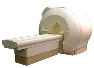
'Next generation MRI system 1.5T CHORUS developed by ISOL Technology is optimized for both clinical diagnostic imaging and for research development.
CHORUS offers the complete range of feature oriented advanced imaging techniques- for both clinical routine and research. The compact short bore magnet, the patient friendly design and the gradient technology make the innovation to new degree of perfection in magnetic resonance.'
Device Information and Specification
CLINICAL APPLICATION
Whole body
Spin Echo, Gradient Echo, Fast Spin Echo,
Inversion Recovery ( STIR, Fluid Attenuated Inversion Recovery), FLASH, FISP, PSIF, Turbo Flash ( MPRAGE ),TOF MR Angiography, Standard echo planar imaging package (SE-EPI, GE-EPI), Optional:
Advanced P.A. Imaging Package (up to 4 ch.), Advanced echo planar imaging package,
Single Shot and Diffusion Weighted EPI, IR/FLAIR EPI
STRENGTH
20 mT/m (Upto 27 mT/m)
| |  | |
• View the DATABASE results for 'CHORUS 1.5T™' (2).
| | | | |
|  | |  |  |
|  | |
|  | | |
|
| |
 | Look
Ups |
| |