Magnetic Resonance - Technology Information Portal Welcome to MRI Technology
Info
  Sheets

Out-
      side


 'Sequences' 
SEARCH FOR    
 
  2 3 5 A B C D E F G H I J K L M N O P Q R S T U V W X Y Z
Result : Searchterm 'Sequences' found in 2 terms [] and 188 definitions []
previous     16 - 20 (of 190)     next
Result Pages : [1]  [2 3 4 5 6 7 8 9 10 11 12 13 14 15 16 17 18 19 20 ... ]
Searchterm 'Sequences' was also found in the following services: 
spacer
News  (6)  Resources  (8)  Forum  (51)  
 
Flip Angle
 
(FA) The flip angle a is used to define the angle of excitation for a field echo pulse sequence. It is the angle to which the net magnetization is rotated or tipped relative to the main magnetic field direction via the application of a RF excitation pulse at the Larmor frequency. It is also referred to as the tip angle, nutation angle or angle of nutation.
The radio frequency power (which is proportional to the square of the amplitude) of the pulse is proportional to a through which the spins are tilted under its influence. Flip angles between 0° and 90° are typically used in gradient echo sequences, 90° and a series of 180° pulses in spin echo sequences and an initial 180° pulse followed by a 90° and a 180° pulse in inversion recovery sequences.
spacer
 
• Related Searches:
    • Precess
    • Pulse, 90°
    • Echo
    • Magnetic Resonance
    • Out of Phase
 
Further Reading:
  Basics:
What MRI Sequences Produce the Highest Specific Absorption Rate (SAR), and Is There Something We Should Be Doing to Reduce the SAR During Standard Examinations?
Thursday, 16 April 2015   by www.ajronline.org    
Mapping of low flip angles in magnetic resonance(.pdf)
Saturday, 1 January 2011   by www.hal.inserm.fr    
  News & More:
A practical guideline for T1 reconstruction from various flip angles in MRI
Saturday, 1 October 2016   by journals.sagepub.com    
Clinical evaluation of a speed optimized T2 weighted fast spin echo sequence at 3.0 T using variable flip angle refocusing, half-Fourier acquisition and parallel imaging
Wednesday, 25 October 2006
Searchterm 'Sequences' was also found in the following services: 
spacer
Radiology  (2) Open this link in a new windowUltrasound  (1) Open this link in a new window
Flow CompensationInfoSheet: - Artifacts - 
Case Studies, 
Reduction Index, 
etc.
 
Flow compensation is based on the principle of even echo rephasing and a function of specific pulse sequences, wherein the application of strategic gradient pulses can compensate for the objectionable spin phase effects of flow motion. Gradient moment nulling of the first order of flow is another adjustment for the reduction of flow artifacts.
Gradient field changes can be configured in such a way that during an echo the magnetization signal vectors for all pixels have zero phase angle independent of velocities, accelerations etc. of the measured tissue. The simplest velocity-compensated pulse sequence is the symmetrical second echo of a spin echo pulse sequence.
Strategic gradient pulses are integrated in special sequences (e.g. CRISP, Complex Rephasing Integrated with Surface Probes) and for the most sequences flow compensation is an optional parameter.
spacer

• View the DATABASE results for 'Flow Compensation' (14).Open this link in a new window

 
Further Reading:
  Basics:
Motion Compensation in MR Imaging
   by ccn.ucla.edu    
Flow comp off: An easy technique to confirm CSF flow within syrinx and aqueduct
Wednesday, 2 January 2013   by medind.nic.in    
MRI Resources 
Chemistry - Pathology - Abdominal Imaging - Bioinformatics - Service and Support - Contrast Agents
 
Spin Echo SequenceInfoSheet: - Sequences - 
Intro, 
Overview, 
Types of, 
etc.MRI Resource Directory:
 - Sequences -
 
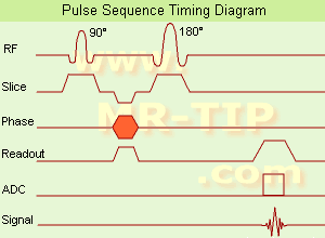
Spin Echo Timing Diagram (SE) The most common pulse sequence used in MR imaging is based of the detection of a spin or Hahn echo. It uses 90° radio frequency pulses to excite the magnetization and one or more 180° pulses to refocus the spins to generate signal echoes named spin echoes (SE).
In the pulse sequence timing diagram, the simplest form of a spin echo sequence is illustrated.
The 90° excitation pulse rotates the longitudinal magnetization (Mz) into the xy-plane and the dephasing of the transverse magnetization (Mxy) starts.
The following application of a 180° refocusing pulse (rotates the magnetization in the x-plane) generates signal echoes. The purpose of the 180° pulse is to rephase the spins, causing them to regain coherence and thereby to recover transverse magnetization, producing a spin echo.
The recovery of the z-magnetization occurs with the T1 relaxation time and typically at a much slower rate than the T2-decay, because in general T1 is greater than T2 for living tissues and is in the range of 100-2000 ms.
The SE pulse sequence was devised in the early days of NMR days by Carr and Purcell and exists now in many forms: the multi echo pulse sequence using single or multislice acquisition, the fast spin echo (FSE/TSE) pulse sequence, echo planar imaging (EPI) pulse sequence and the gradient and spin echo (GRASE) pulse sequence;; all are basically spin echo sequences.
In the simplest form of SE imaging, the pulse sequence has to be repeated as many times as the image has lines.
Contrast values:
PD weighted: Short TE (20 ms) and long TR.
T1 weighted: Short TE (10-20 ms) and short TR (300-600 ms)
T2 weighted: Long TE (greater than 60 ms) and long TR (greater than 1600 ms)
With spin echo imaging no T2* occurs, caused by the 180° refocusing pulse. For this reason, spin echo sequences are more robust against e.g., susceptibility artifacts than gradient echo sequences.

See also Pulse Sequence Timing Diagram to find a description of the components.
 
Images, Movies, Sliders:
 Shoulder Coronal T1 SE  Open this link in a new window
    

Courtesy of  Robert R. Edelman
 Shoulder Axial T1 SE  Open this link in a new window
 MRI Orbita T1  Open this link in a new window
    
 
spacer

• View the DATABASE results for 'Spin Echo Sequence' (24).Open this link in a new window

 
Further Reading:
  Basics:
Fast Spin Echo(.pdf)
Tuesday, 24 January 2006   by www.81bones.net    
Magnetic resonance imaging
   by www.scholarpedia.org    
FUNDAMENTALS OF MRI: Part I
   by www.e-radiography.net    
  News & More:
New MR sequence helps radiologists more accurately evaluate abnormalities of the uterus and ovaries
Thursday, 23 April 2009   by www.eurekalert.org    
MRI techniques improve pulmonary embolism detection
Monday, 19 March 2012   by medicalxpress.com    
Searchterm 'Sequences' was also found in the following services: 
spacer
News  (6)  Resources  (8)  Forum  (51)  
 
Spoiled Gradient Echo SequenceInfoSheet: - Sequences - 
Intro, 
Overview, 
Types of, 
etc.MRI Resource Directory:
 - Sequences -
 
Spoiled gradient echo sequences use a spoiler gradient on the slice select axis during the end module to destroy any remaining transverse magnetization after the readout gradient, which is the case for short repetition times.
As a result, only z-magnetization remains during a subsequent excitation. This types of sequences use semi-random changes in the phase of radio frequency pulses to produce a spatially independent phase shift.
Companies use different acronyms to describe certain techniques.

Different terms for these gradient echo pulse sequences:
CE-FFE-T1 Contrast Enhanced Fast Field Echo with T1 Weighting,
GFE Gradient Field Echo,
FLASH Fast Low Angle Shot,
PS Partial Saturation,
RF spoiled FAST RF Spoiled Fourier Acquired Steady State Technique,
RSSARGE Radio Frequency Spoiled Steady State Acquisition Rewound Gradient Echo
S-GRE Spoiled Gradient Echo,
SHORT Short Repetition Techniques,
SPGR Spoiled Gradient Recalled (spoiled GRASS),
STAGE T1W T1 weighted Small Tip Angle Gradient Echo,
T1-FAST T1 weighted Fourier Acquired Steady State Technique,
T1-FFE T1 weighted Fast Field Echo.
In this context, 'contrast enhanced' refers to the pulse sequence, it does not mean enhancement with a contrast agent.
spacer

• View the DATABASE results for 'Spoiled Gradient Echo Sequence' (11).Open this link in a new window

 
Further Reading:
  News & More:
3-D VOLUMETRIC IMAGING FOR STEREOTACTIC LESIONAL AND DEEP BRAIN STIMULATION SURGERY
Cutting Edge Imaging of THE Spine
February 2007   by www.pubmedcentral.nih.gov    
Searchterm 'Sequences' was also found in the following services: 
spacer
Radiology  (2) Open this link in a new windowUltrasound  (1) Open this link in a new window
Black Boundary ArtifactInfoSheet: - Artifacts - 
Case Studies, 
Reduction Index, 
etc.MRI Resource Directory:
 - Artifacts -
 
Quick Overview
Please note that there are different common names for this artifact.
Artifact Information
NAME
Black boundary, dark boundary, contour, chemical shift, relief
DESCRIPTION
Black contours at boundaries
Black boundary artifacts are black lines following voxels where both water and fat protons are present in the same voxel. This artifact arise along the boundary of organs or tissues perpendicular to the frequency encoding direction, and occurs preferentially in gradient echo sequences with out of phase echo times.
mri safety guidance
Image Guidance
Fat suppression techniques eliminate this artifact. For artifact reducing helps a smaller water fat shift (high bandwidth), a higher matrix or/and an in phase TE.

See also Chemical Shift Artifact.
spacer

• View the DATABASE results for 'Black Boundary Artifact' (4).Open this link in a new window

 
Further Reading:
  Basics:
What is chemical shift artefact? Why does it occur? How many Hz at 1.5 T?
   by www.revisemri.com    
MRI Resources 
Implant and Prosthesis - Devices - PACS - Pediatric and Fetal MRI - Sequences - MRI Centers
 
previous      16 - 20 (of 190)     next
Result Pages : [1]  [2 3 4 5 6 7 8 9 10 11 12 13 14 15 16 17 18 19 20 ... ]
 Random Page
 
Share This Page
FacebookTwitterLinkedIn

MR-TIP    
Community   
User
Pass
Forgot your UserID/Password ?    


MRI is trending to low field magnets :
reduced costs will lead to this change 
AI will close the gap to high field 
only in remote areas 
is only temporary 
never 

Look
      Ups


MR-TIP.com uses cookies! By browsing MR-TIP.com, you agree to our use of cookies.

Magnetic Resonance - Technology Information Portal
Member of SoftWays' Medical Imaging Group - MR-TIP • Radiology-TIP • Medical-Ultrasound-Imaging • 
Copyright © 2003 - 2024 SoftWays. All rights reserved. [ 21 January 2025]
Terms of Use | Privacy Policy | Advertising
 [last update: 2024-02-26 03:41:00]