 | Info
Sheets |
| | | | | | | | | | | | | | | | | | | | | | | | |
 | Out-
side |
| | | | |
|
| | | | |
Result : Searchterm 'Slab' found in 3 terms [ ] and 24 definitions [ ] and 24 definitions [ ] ]
| previous 6 - 10 (of 27) nextResult Pages :  [1] [1]  [2 3 4 5 6] [2 3 4 5 6] |  | |  | Searchterm 'Slab' was also found in the following services: | | | | |
|  |  |
| |
|
(3D MRA) The 3D angiography technique can be applied to focus on fast flowing (arterial) blood and to visualize small tortuous vessels. 3D TOF images are less sensitive to turbulent flow artifacts.
The advantage of this approach is that the signal, acquired from the entire
volume has an increased signal to noise ratio. Slices are defined by a second phase encoded axis, which divides the volume into 'partitions'.
3D TOF MRA is acquired with 3D FT slabs or multiple overlapping thin 3D FT slabs ( MOTSA) depending on the coverage required and the range of flow-velocities under examination.
Such 3D techniques can provide equal spatial resolution along all three axes, i.e. be 'isotropic', or the partition thickness can be greater or less than the in plane spatial resolution in which case can be said to be 'anisotropic'.
The circle of Willis, anatomy as well as its fast arterial flow, lends itself well to both 3D TOF and 2D or 3D phase contrast angiography. | | | |  | | | | | | | | |  Further Reading: Further Reading: | Basics:
|
|
| |
|  |  | Searchterm 'Slab' was also found in the following service: | | | | |
|  |  |
| |
|
A technique, which produces a 3 dimensional image of an object. The advantage of this approach is that the signal, acquired from the entire volume has an increased SNR. 'Slices' are defined by a second phase encoded axis, which divides the volume into 'partitions'.
There is no gap between the slices in 3D volume imaging, therefore thin slices are possible. The Gz phase encoding gradient is set for several slices in one. But 3D takes more time with thin slices because of this phase encoding gradient. With conventional thin slice imaging, the SNR is poor, with 3D volume imaging this is not the case because the slab (volume) is responsible for SNR. | | | |  | |
• View the DATABASE results for '3 Dimensional Imaging' (5).
| | |
• View the NEWS results for '3 Dimensional Imaging' (1).
| | | | |  Further Reading: Further Reading: | | Basics:
|
|
News & More:
| |
| |
|  | |  |  |  |
| |
|

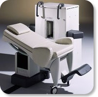
Developed by GE Lunar; the ARTOSCAN™-M is designed specifically for in-office musculoskeletal imaging. ARTOSCAN-M's compact, modular design allows placing within a clinical environment, bringing MRI to the patient. Patients remain outside the magnet at all times during the examinations, enabling constant patient-technologist contact. ARTOSCAN-M requires no special RF room, magnetic shielding, special power supply or air conditioning.
The C-SCAN™ (also known as Artoscan C) is developed from the ARTOSCAN™ - M, with a new computer platform.
Device Information and Specification
CLINICAL APPLICATION
Dedicated extremity
SE, GE, IR, STIR, FSE, 3D CE, GE-STIR, 3D GE, ME, TME, HSE
SLICE THICKNESS
2D: 2 mm - 10 mm;
3D: 0.6 mm - 10 mm
4,096 gray lvls, 256 lvls in 3D
POWER REQUIREMENTS
100/110/200/220/230/240V
| |  | |
• View the DATABASE results for 'ARTOSCAN™ - M' (3).
| | | | |
|  |  | Searchterm 'Slab' was also found in the following services: | | | | |
|  |  |
| |
|
Contact Information
MAIL
Bornhop Research Group
MS 1061 Dept of Chemistry and Biochemistry
Texas Tech University
Lubbock, TX 79409
USA
PHONE
Office Phone: +1-806-742-3142
Lab Phone: +1-806-742-3152
| |  | | | |
|  |  | Searchterm 'Slab' was also found in the following service: | | | | |
|  |  |
| |
|
Device Information and Specification
CLINICAL APPLICATION
Dedicated extremity
SE, GE, IR, STIR, FSE, 3D CE, GE-STIR, 3D GE, ME, TME, HSE
IMAGING MODES
Single, multislice, volume study, fast scan, multi slab
2D: 2 mm - 10 mm;
3D: 0.6 mm - 10 mm
4,096 gray lvls, 256 lvls in 3D
POWER REQUIREMENTS
100/110/200/220/230/240
| |  | |
• View the DATABASE results for 'C-SCAN™' (4).
| | | | |
|  | |  |  |
|  | | |
|
| |
 | Look
Ups |
| |