Magnetic Resonance - Technology Information Portal Welcome to MRI Technology
Info
  Sheets

Out-
      side


 'Slew Rate' 
SEARCH FOR    
 
  2 3 5 A B C D E F G H I J K L M N O P Q R S T U V W X Y Z
Result : Searchterm 'Slew Rate' found in 1 term [] and 15 definitions []
1 - 5 (of 16)     next
Result Pages : [1]  [2 3 4]
Searchterm 'Slew Rate' was also found in the following service: 
spacer
Forum  (9)  
 
Slew RateForum -
related threads
 
The gradient slew rate is the speed rate of ascent or descent of a gradient from zero to its maximum amplitude, either positive or negative. Which is the amplitude divided by the rise time in msec. Measured in mT/m/msec or T/m/sec. The shorter the rise time, the faster the gradients and therefore echo spacing. Gradients with a shorter echo spacing will have a better resolution and more slices per TR.
spacer
 
• Share the entry 'Slew Rate':  Facebook  Twitter  LinkedIn  
 
• Related Searches:
    • Magnetic Field Gradient
    • Rise Time
    • Tesla
    • Echo Time
    • Echo Train
MRI Resources 
Implant and Prosthesis - Liver Imaging - DICOM - MRI Training Courses - MRI Technician and Technologist Jobs - Pediatric and Fetal MRI
 
Echelon™ 1.5TInfoSheet: - Devices -
Intro, 
Types of Magnets, 
Overview, 
etc.
 
www.hitachimed.com/contentindex.asp?ID=971 From Hitachi Medical Systems America Inc.;
Hitachi expanded its portfolio with the Echelon™ 1.5T. The MRI scanner combines a compact magnet and a scalable 8-channel RF system with high-performance gradients and slew rate to select short echo times, small field of views, high matrices and thin slices. Standard features of the Echelon MRI system include higher-order active shim, RAPID (parallel imaging for use on brain MRI, body, cardiovascular imaging, and orthopedic coils), multiple coil ports, and an advanced reconstruction engine.
Device Information and Specification
CLINICAL APPLICATION
Whole body
CONFIGURATION
Short bore
Head, body coil, spine, breast, knee, shoulder, vascular multiple array coils.
SYNCHRONIZATION
Cardiac gating, ECG/peripheral, respiratory gating
PULSE SEQUENCES
SE, IR, FSE, FIR, GE, SG, BASG, PBSG, PCIR, DWI, Radial, Angiography: TOF, FLUTE (Fluoro-triggered bolus MRA), Time-resolved MRA
IMAGING MODES
Single, multislice, volume study
PIXEL INTENSITY
Level Range: -2,000 to +4,000
Sub millimeter
POWER REQUIREMENTS
208/220/240 V, single phase
CRYOGEN USE
Low cryogen boil-off
STRENGTH
30 mT/m
150 T/m/sec
Higher-order active shim
spacer

• View the DATABASE results for 'Echelon™ 1.5T' (2).Open this link in a new window


• View the NEWS results for 'Echelon™ 1.5T' (3).Open this link in a new window.
 
Further Reading:
  Basics:
Echelon 1.5T
   by www.hitachimed.com    
MRI Resources 
MRI Technician and Technologist Career - Stent - Mobile MRI Rental - Spectroscopy pool - Functional MRI - Sequences
 
Biograph mMR
 
www.healthcare.siemens.com/magnetic-resonance-imaging/mr-pet-scanner/biograph-mmr FDA cleared and CE Mark 2011.
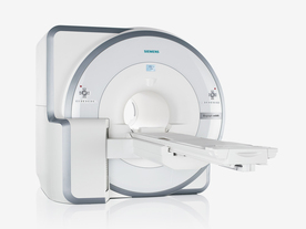
The Biograph mMR has a fully-integrated design for simultaneous PET/MRI imaging. The dedicated hardware includes solid-state, avalanche photodiode PET detector and adapted, PET-compatible MR coils.
The possibility of truly simultaneous operation allows the acquisition of several magnetic resonance imaging (MRI) sequences during the positron emission tomography (PET) scan, without increasing the examination time.
See also Hybrid Imaging.
Device Information and Specification
CLINICAL APPLICATION
Whole Body
CONFIGURATION
Simultaneous PET/MRI
3 Tesla
Full range of MRI coils
CHANNELS
up to 32
PET ACQUISITION MODES
3D
MRI ACQUISITION MODES
2D/3D, spectroscopy;; iPAT, GRAPPA (k-space), noncontrast angiography, radial motion compensation
PET EFFECTIVE AXIAL FOV
26 cm (typical overlap 23%)
MRI FOV
A-P 45, R-L 50, H-F 50 cm
PET RING DIAMETER
65.6 cm
PATIENT SCAN RANGE
199 cm
HORIZONTAL SPEED
200 mmsec
TABLE CAPACITY
200 kg
PET DETECTOR
Solid state, 4032 avalanche photo diodes
DETECTOR SCINTILLATION MATERIAL
LSO, 28672 crystals
CRYSTAL SIZE
4 x 4 x 20 mm
MAGNET WEIGHT
9000 kg
DIMENSION H*W*D (gantry included)
335 x 230 x 242 cm (finshed covers)
5-GAUSS FRINGE FIELD
2.6 m / 4.6 m
CRYOGEN USE
Zero boil off rate, approx. 10 years
COOLING SYSTEM
PET system: water; MRI system: water
up to 200 T/m/s
MAX. AMPLITUDE
45 mT/m
Aautomatic, patient specific shim; active shim 3 linear and 5 non-linear channels (seond order)
POWER REQUIREMENTS
380 / 400 / 420 / 440 / 460 / 480 V, 3-phase + ground; Total system 110kW
spacer
 
Further Reading:
  Basics:
Performance Measurements of the Siemens mMR Integrated Whole-Body PET/MR Scanner
Friday, 11 November 2011   by jnm.snmjournals.org    
Global Trends in Hybrid Imaging
Monday, 1 November 2010   by pubs.rsna.org    
  News & More:
Positron Emission Tomographic Imaging in Stroke
Monday, 28 December 2015   by www.ncbi.nlm.nih.gov    
A world's first in imaging – integrated whole-body molecular MR system available for clinical use testing
Thursday, 18 November 2010   by www.siemens.com    
FDA Clears New System to Perform Simultaneous PET, MRI Scans
Friday, 10 June 2011   by www.prnewswire.com    
Frost & Sullivan Award Underlines Siemens Healthcare's Contribution to New Product Innovation in the North American Medical Imaging Market
Wednesday, 13 July 2011   by multivu.prnewswire.com    
Searchterm 'Slew Rate' was also found in the following service: 
spacer
Forum  (9)  
 
Coronary AngiographyMRI Resource Directory:
 - Cardiovascular Imaging -
 
(MRI-CA, MRCA) The noninvasive imaging of the coronary arteries using magnetic resonance imaging of the heart.
For cardiac MRI-CA, high performance machines are necessary with minimum 40mT/m and 300μsec slew rate.
2D and 3D acquisition are used for fast gradient echo sequences with techniques for minimizing cardiac and respiratory motion and suppressing the high signal of pericardial fat. The optimal sequences seem to be trueFISP, Balanced FFE or FIESTA with SMASH and SENSE techniques. Respiratory motion is minimized for 3D acquisitions by using respiratory gating, especially using navigator echoes (Navigator Technique) to track diaphragmatic and cardiac movement. Optimization of MR technique can provide mapping of long segments of the coronary arteries.
Blood pool agents are being applied to improve the reliability of coronary MR angiography. The major current clinical indication is the identification of coronary artery anomalies because the diagnostic accuracy's for identifying haemodynamically significant stenoses are variable depending of the image quality.

See also Magnetic Resonance Angiography, and Cardiac MRI.
spacer

• View the DATABASE results for 'Coronary Angiography' (7).Open this link in a new window

 
Further Reading:
  Basics:
Role of Magnetic Resonance Imaging in Visualizing Coronary Arteries
Monday, 2 August 2004   by www.clinmedres.org    
  News & More:
Graphic illustration
Tuesday, 12 February 2008   by www.theengineer.co.uk    
MRI Resources 
Abdominal Imaging - Research Labs - Education pool - Devices - Lung Imaging - Knee MRI
 
GradientForum -
related threads
 
The word gradient (from grade) means the inclination of a surface along a given direction. In MRI, gradient stands for gradient field and/or gradient coil. Inside the main magnet are three gradient coils located, which produce the desired gradient (magnetic) fields. These fields are used to alter (collectively and sequentially) the influence of the static magnetic field B0 on the imaged object by inc- or decreasing the field strength and changing the direction. Through this influence selective spatial excitation and spatial encoding (each voxel resonate at a different frequency) is possible. Gradients are also utilized in another way for fast imaging sequences.

See also Slew Rate and Duty Cycle.
spacer

• View the DATABASE results for 'Gradient' (316).Open this link in a new window


• View the NEWS results for 'Gradient' (2).Open this link in a new window.
 
Further Reading:
  Basics:
Active Noise Control of Magnetic Resonance Imaging Scanner Using Inverse Modeling Technique
Monday, 19 April 2010   by www.acoustics.org    
  News & More:
On the Horizon - Next Generation MRI
Wednesday, 23 October 2013   by thefutureofthings.com    
MRI Resources 
Veterinary MRI - Corporations - Pathology - PACS - Brain MRI - DICOM
 
     1 - 5 (of 16)     next
Result Pages : [1]  [2 3 4]
 Random Page
 
Share This Page
FacebookTwitterLinkedIn

MR-TIP    
Community   
User
Pass
Forgot your UserID/Password ?    


How AI will impact MRI :
only diagnostics 
saving time 
reducing cost 
makes planning obsolete 
reduce human knowledge 
not at all 

Look
      Ups


MR-TIP.com uses cookies! By browsing MR-TIP.com, you agree to our use of cookies.

Magnetic Resonance - Technology Information Portal
Member of SoftWays' Medical Imaging Group - MR-TIP • Radiology-TIP • Medical-Ultrasound-Imaging • 
Copyright © 2003 - 2024 SoftWays. All rights reserved. [ 21 November 2024]
Terms of Use | Privacy Policy | Advertising
 [last update: 2024-02-26 03:41:00]