 | Info
Sheets |
| | | | | | | | | | | | | | | | | | | | | | | | |
 | Out-
side |
| | | | |
|
| | | | | |  | Searchterm 'Slice' was also found in the following services: | | | | |
|  |  |
| |
|

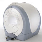
From GE Healthcare;
The Signa HDx MRI system is GE's leading edge whole body magnetic resonance scanner designed to support high resolution, high signal to noise ratio, and short scan times.
Signa HDx 3.0T offers new technologies like ultra-fast image reconstruction through the new XVRE recon engine, advancements in parallel imaging algorithms and the broadest range of premium applications. The HD applications, PROPELLER (high-quality brain imaging extremely resistant to motion artifacts), TRICKS (contrast-enhanced angiographic vascular lower leg imaging), VIBRANT (for breast MRI), LAVA (high resolution liver imaging with shorter breath holds and better organ coverage) and MR Echo (high-definition cardiac images in real time) offer unique capabilities.
Device Information and Specification CLINICAL APPLICATION Whole body
CONFIGURATION Compact short bore SE, IR, 2D/3D GRE, RF-spoiled GRE, 2DFGRE, 2DFSPGR, 3DFGRE, 3DFSPGR, 3DTOFGRE, 3DFSPGR, 2DFSE, 2DFSE-XL, 2DFSE-IR, T1-FLAIR, SSFSE, EPI, DW-EPI, BRAVO, Angiography: 2D/3D TOF, 2D/3D phase contrast vascular IMAGING MODES Single, multi slice, volume study, fast scan, multi slab, cine, localizer H*W*D 240 x 2216,6 x 201,6 cm POWER REQUIREMENTS 480 or 380/415, 3 phase ||
COOLING SYSTEM TYPE Closed-loop water-cooled grad. | |  | | | |
|  |  | Searchterm 'Slice' was also found in the following services: | | | | |
|  |  |
| |
|
| | | |  | |
• View the DATABASE results for 'Spin Phase Effect' (3).
| | | | |
|  | |  |  |  |
| |
|
A collection of slices in a multi slice acquisition with the same orientation. Multi-stack acquisitions are possible when the stacks parameter is greater than one. | |  | |
• View the DATABASE results for 'Stacks' (4).
| | | | |
|  |  | Searchterm 'Slice' was also found in the following services: | | | | |
|  |  |
| |
|
Tomography is imaging by sections or sectioning. A device used in tomography is called a tomograph, while the image produced is a tomogram.
The mathematical basis for tomographic imaging was laid down by Johann Radon. It is applied in computed tomography and magnetic resonance tomography (MRT) also called magnetic resonance imaging ( MRI) to obtain cross-sectional images of slices through the body of patients. Each of that slices is defined by thickness and spatial resolution (see voxel). | | | |  | |
• View the DATABASE results for 'Tomographic Imaging' (9).
| | | | |  Further Reading: Further Reading: | | Basics:
|
|
News & More:
| |
| |
|  |  | Searchterm 'Slice' was also found in the following services: | | | | |
|  |  |
| |
|
Device Information and Specification
CLINICAL APPLICATION
Whole body
CONFIGURATION
Mobile compact
Whole body, intra-operative head, neck volume, atlas head//neck vascular quadrature phased array, spine quadrature, C/T/L spine phased array, small joint, large joint, TMJ bilateral, shoulder phased array, extremity quadrature volume, wrist, hand quadrature, general purpose flexible, pelvis/abdomen phased array, body quadrature, phased array flexible, breast bilateral
IMAGING MODES
Localizer, single slice, multi slice, volume
| |  | |
• View the DATABASE results for 'iMotion™ 1.5 Tesla Magnet' (2).
| | | | |
|  | |  |  |
|  | |
|  | | |
|
| |
 | Look
Ups |
| |