 | Info
Sheets |
| | | | | | | | | | | | | | | | | | | | | | | | |
 | Out-
side |
| | | | |
|
| | | | |
Result : Searchterm 'Slice Thickness' found in 1 term [ ] and 63 definitions [ ] and 63 definitions [ ] ]
| previous 6 - 10 (of 64) nextResult Pages :  [1] [1]  [2 3 4 5 6 7 8 9 10 11 12 13] [2 3 4 5 6 7 8 9 10 11 12 13] |  | |  | Searchterm 'Slice Thickness' was also found in the following services: | | | | |
|  |  |
| |
|

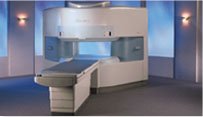
From Hitachi Medical Systems America, Inc.;
the AIRIS made its debut in 1995. Hitachi followed up with the AIRIS II system, which has proven equally successfully. 'All told, Hitachi has installed more than 1,000 MRI systems in the U.S., holding more than 17 percent of the total U.S. MRI installed base, and more than half of the installed base of open MR systems,' says Antonio Garcia, Frost and Sullivan industry research analyst.
Now Altaire employs a blend of innovative Hitachi features called VOSIā¢ technology, optimizing each sub-system's performance in concert with the
other sub-systems, to give the seamless mix of high-field performance
and the patient comfort, especially for claustrophobic patients, of open MR systems.
Device Information and Specification
CLINICAL APPLICATION
Whole body
DualQuad T/R Body Coil, MA Head, MA C-Spine, MA Shoulder, MA Wrist, MA CTL Spine, MA Knee, MA TMJ, MA Flex Body (3 sizes), Neck, small and large Extremity, PVA (WIP), Breast (WIP), Neurovascular (WIP), Cardiac (WIP) and MA Foot//Ankle (WIP)
SE, GE, GR, IR, FIR, STIR, ss-FSE, FSE, DE-FSE/FIR, FLAIR, ss/ms-EPI, ss/ms EPI- DWI, SSP, MTC, SE/GE-EPI, MRCP, SARGE, RSSG, TRSG, BASG, Angiography: CE, PC, 2D/3D TOF
IMAGING MODES
Single, multislice, volume study
TR
SE: 30 - 10,000msec GE: 3.6 - 10,000msec IR: 50 - 16,700msec FSE: 200 - 16,7000msec
TE
SE : 8 - 250msec IR: 5.2 -7,680msec GE: 1.8 - 2,000 msec FSE: 5.2 - 7,680
0.05 sec/image (256 x 256)
2D: 2 - 100 mm; 3D: 0.5 - 5 mm
Level Range: -2,000 to +4,000
COOLING SYSTEM TYPE
Water-cooled
3.1 m lateral, 3.6 m vertical
| |  | | | | | | | | |  Further Reading: Further Reading: | News & More:
|
|
| |
|  |  | Searchterm 'Slice Thickness' was also found in the following service: | | | | |
|  |  |
| |
|
A description of the factors used in creating an image should
include the type and times of the pulse sequence, the number of signals averaged or added ( NSA), the size of the reconstructed region, the size of
the acquisition matrix in each direction, field of view and the slice thickness; usually printed at the border of MRI pictures. | |  | | | |
|  | |  |  |  |
| |
|
Device Information and Specification
CLINICAL APPLICATION
Dedicated extremity
SE, GE, IR, STIR, FSE, 3D CE, GE-STIR, 3D GE, ME, TME, HSE
IMAGING MODES
Single, multislice, volume study, fast scan, multi slab
2D: 2 mm - 10 mm;
3D: 0.6 mm - 10 mm
4,096 gray lvls, 256 lvls in 3D
POWER REQUIREMENTS
100/110/200/220/230/240
| |  | |
• View the DATABASE results for 'C-SCAN™' (4).
| | | | |
|  |  | Searchterm 'Slice Thickness' was also found in the following services: | | | | |
|  |  |
| |
|
Cervical spine MRI is a suitable tool in the assessment of all cervical spine (vertebrae C1 - C7) segments (computed tomography (CT) images may be unsatisfactory close to the thoracic spine due to shoulder artifacts). The cervical spine is particularly susceptible to degenerative problems caused by the complex anatomy and its large range of motion.
Advantages of magnetic resonance imaging MRI are the high soft tissue contrast (particularly important in diagnostics of the spinal cord), the ability to display the entire spine in sagittal views and the capacity of 3D visualization. Magnetic resonance myelography is a useful supplement to conventional MRI examinations in the investigation of cervical stenosis. Myelographic sequences result in MR images with high contrast that are similar in appearance to conventional myelograms. Additionally, open MRI studies provide the possibility of weight-bearing MRI scan to evaluate structural positional and kinetic changes of the cervical spine. Indications of cervical spine MRI scans include the assessment of soft disc herniations, suspicion of disc hernia recurrence after operation, cervical spondylosis, osteophytes, joint arthrosis, spinal canal lesions (tumors, multiple sclerosis, etc.), bone diseases (infection, inflammation, tumoral infiltration) and paravertebral spaces.
State-of-the-art phased array spine coils and high performance MRI machines provide high image quality and short scan time. Imaging protocols for the cervical spine includes sagittal T1 weighted and T2 weighted sequences with 3-4 mm slice thickness and axial slices; usually contiguous from C2 through T1. Additionally, T2 fat suppressed and T1 post contrast images are often useful in spine imaging. See also Lumbar Spine MRI.
| |  | |
• View the DATABASE results for 'Cervical Spine MRI' (2).
| | |
• View the NEWS results for 'Cervical Spine MRI' (1).
| | | | |  Further Reading: Further Reading: | News & More:
|
|
| |
|  |  | Searchterm 'Slice Thickness' was also found in the following service: | | | | |
|  |  |
| |
|
(DQA) This MRI scan or MRI procedure is used by system operators to verify system operation based on relevant image quality parameters like e.g., SNR, slice thickness, geometric distortion, slice position, image resolution and ghosting.
The quality assurance should carry out according to instructions of the manufacturer, normally using the head coil. In addition, SNR can be measured monthly on a selection of commonly used coils.
Weekly recording of these parameters is recommended for clinical MRI machines, as this allows early detecting of deviations from acceptable limits. | |  | |
• View the DATABASE results for 'Daily Quality Assurance' (3).
| | | | |  Further Reading: Further Reading: | News & More:
|
|
| |
|  | |  |  |
|  | |
|  | | |
|
| |
 | Look
Ups |
| |