 | Info
Sheets |
| | | | | | | | | | | | | | | | | | | | | | | | |
 | Out-
side |
| | | | |
|
| | | | | |  | Searchterm 'Spatial Resolution' was also found in the following services: | | | | |
|  |  |
| |
|

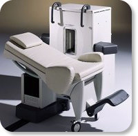
Developed by GE Lunar; the ARTOSCAN™-M is designed specifically for in-office musculoskeletal imaging. ARTOSCAN-M's compact, modular design allows placing within a clinical environment, bringing MRI to the patient. Patients remain outside the magnet at all times during the examinations, enabling constant patient-technologist contact. ARTOSCAN-M requires no special RF room, magnetic shielding, special power supply or air conditioning.
The C-SCAN™ (also known as Artoscan C) is developed from the ARTOSCAN™ - M, with a new computer platform.
Device Information and Specification
CLINICAL APPLICATION
Dedicated extremity
SE, GE, IR, STIR, FSE, 3D CE, GE-STIR, 3D GE, ME, TME, HSE
SLICE THICKNESS
2D: 2 mm - 10 mm;
3D: 0.6 mm - 10 mm
4,096 gray lvls, 256 lvls in 3D
POWER REQUIREMENTS
100/110/200/220/230/240V
| |  | | | | | | | | |
|  |  | Searchterm 'Spatial Resolution' was also found in the following service: | | | | |
|  |  |
| |
|

From Hitachi Medical Systems America, Inc.;
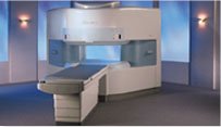
the AIRIS made its debut in 1995. Hitachi followed up with the AIRIS II system, which has proven equally successfully. 'All told, Hitachi has installed more than 1,000 MRI systems in the U.S., holding more than 17 percent of the total U.S. MRI installed base, and more than half of the installed base of open MR systems,' says Antonio Garcia, Frost and Sullivan industry research analyst.
Now Altaire employs a blend of innovative Hitachi features called VOSI™ technology, optimizing each sub-system's performance in concert with the
other sub-systems, to give the seamless mix of high-field performance
and the patient comfort, especially for claustrophobic patients, of open MR systems.
Device Information and Specification
CLINICAL APPLICATION
Whole body
DualQuad T/R Body Coil, MA Head, MA C-Spine, MA Shoulder, MA Wrist, MA CTL Spine, MA Knee, MA TMJ, MA Flex Body (3 sizes), Neck, small and large Extremity, PVA (WIP), Breast (WIP), Neurovascular (WIP), Cardiac (WIP) and MA Foot//Ankle (WIP)
SE, GE, GR, IR, FIR, STIR, ss-FSE, FSE, DE-FSE/FIR, FLAIR, ss/ms-EPI, ss/ms EPI- DWI, SSP, MTC, SE/GE-EPI, MRCP, SARGE, RSSG, TRSG, BASG, Angiography: CE, PC, 2D/3D TOF
IMAGING MODES
Single, multislice, volume study
TR
SE: 30 - 10,000msec GE: 3.6 - 10,000msec IR: 50 - 16,700msec FSE: 200 - 16,7000msec
TE
SE : 8 - 250msec IR: 5.2 -7,680msec GE: 1.8 - 2,000 msec FSE: 5.2 - 7,680
0.05 sec/image (256 x 256)
2D: 2 - 100 mm; 3D: 0.5 - 5 mm
Level Range: -2,000 to +4,000
COOLING SYSTEM TYPE
Water-cooled
3.1 m lateral, 3.6 m vertical
| |  | |
• View the DATABASE results for 'Altaire™' (2).
| | | | |  Further Reading: Further Reading: | News & More:
|
|
| |
|  | |  |  | |  |  | Searchterm 'Spatial Resolution' was also found in the following services: | | | | |
|  |  |
| |
|
( BOLD) In MRI the changes in blood oxygenation level are visible. Oxyhaemoglobin (the principal haemoglobin in arterial blood) has no substantial magnetic properties, but deoxyhaemoglobin (present in the draining veins after the oxygen has been unloaded in the tissues) is strongly paramagnetic. It can thus serve as an intrinsic paramagnetic contrast agent in appropriately performed brain MRI. The concentration and relaxation properties of deoxyhaemoglobin make it a susceptibility , e.g. T2 relaxation effective contrast agent with little effect on T1 relaxation.
During activation of the brain, the oxygen consumption of the local tissue increase by approximately 5% with that the oxygen tension will decrease. As a consequence, after a short period of time vasodilatation occurs, resulting in a local increase of blood volume and flow by 20 - 40%. The incommensurate change in local blood flow and oxygen extraction increases the local oxygen level.
By using T2 weighted gradient echo EPI sequences, which are highly susceptibility sensitive and fast enough to capture the three-dimensional nature of activated brain areas will show an increase in signal intensity as oxyhaemoglobin is diamagnetic and deoxyhaemoglobin is paramagnetic. Other MR pulse sequences, such as spoiled gradient echo pulse sequences are also used.
As the effects are subtle and of the order of 2% in 1.5 T MR imaging, sophisticated methodology, paradigms and data analysis techniques have to be used to consistently demonstrate the effect.
As the BOLD effect is due to the deoxygenated blood in the draining veins, the spatial localization of the region where there is increased blood flow resulting in decreased oxygen extraction is not as precisely defined as the morphological features in MRI. Rather there is a physiological blurring, and is estimated that the linear dimensions of the physiological spatial resolution of the BOLD phenomenon are around 3 mm at best. | |  | |
• View the DATABASE results for 'Blood Oxygenation Level Dependent Contrast' (6).
| | | | |  Further Reading: Further Reading: | | Basics:
|
|
News & More:
| |
| |
|  |  | Searchterm 'Spatial Resolution' was also found in the following service: | | | | |
|  |  |
| |
|
(MR mammography) Magnetic resonance imaging of the breast is particularly useful in evaluation of newly diagnosed breast cancer, in women whose breast tissue is mammographically very dense and for screening in women with a high lifetime risk of breast cancer because of their family history or genetic disposition.
Breast MRI can be performed on all standard whole body magnets at a field strength of 0.5 T - 1.5 Tesla. Powerful gradient strengths over 15 mT/m will help to improve the balance between spatial resolution, scanning speed, and volume coverage. The use of a dedicated bilateral breast coil is obligatory.
Malignant lesions release angiogenic factors that increase local vessel density and vessel permeability. Breast cancer is detectable due to the strong enhancement in dynamic breast imaging that peaks early (about 1-2 min.) after contrast medium injection. If breast cancer is suspected, a breast biopsy may be necessary to secure the diagnosis. See also Magnetic Resonance Imaging MRI, Biopsy and MR Guided Interventions.
Requirements in breast MRI procedures:
•
Both breasts must be measured without gaps.
•
For the best possible detection of enhancement fat signal should be eliminated either by image subtraction or by
spectrally selective fat saturation.
•
Thin slices are necessary to assure absence of partial
volume effects.
•
Imaging should be performed with a spatial
resolution in plane less than 1 mm.
For Ultrasound Imaging (USI) see Breast Ultrasound at Medical-Ultrasound-Imaging.com.
See also the related poll result: ' MRI will have replaced 50% of x-ray exams by' | | | | | | | | | | |
• View the DATABASE results for 'Breast MRI' (13).
| | |
• View the NEWS results for 'Breast MRI' (41).
| | | | |  Further Reading: Further Reading: | | Basics:
|
|
News & More:
|  |
Technology advances in breast cancer screenings lead to early diagnosis
Friday, 6 October 2023 by ksltv.com |  |  |
Are synthetic contrast-enhanced breast MRI images as good as the real thing?
Friday, 18 November 2022 by healthimaging.com |  |  |
Abbreviated breast MRI protocols not as cost-effective as promised, new study shows
Wednesday, 20 July 2022 by healthimaging.com |  |  |
Deep learning poised to improve breast cancer imaging
Thursday, 24 February 2022 by www.eurekalert.org |  |  |
Pre-Operative Breast MRI Can Help Identify Patients Likely to Experience Nipple-Sparing Mastectomy Risks
Wednesday, 7 April 2021 by www.diagnosticimaging.com |  |  |
Breast cancer screening recalls: simple MRI measurement could avoid 30% of biopsies
Monday, 1 March 2021 by www.eurekalert.org |  |  |
A Comparison of Methods for High-Spatial-Resolution Diffusion-weighted Imaging in Breast MRI
Tuesday, 25 August 2020 by pubs.rsna.org |  |  |
Pre-Operative Breast MRI Diagnoses More Cancers in Women with DCIS
Thursday, 9 July 2020 by www.diagnosticimaging.com |  |  |
Breast MRI and tumour biology predict axillary lymph node response to neoadjuvant chemotherapy for breast cancer
Thursday, 26 December 2019 by cancerimagingjournal.biomedcentral.com |  |  |
Breast MRI Coding Gets an Overhaul in 2019
Wednesday, 9 January 2019 by www.aapc.com |  |  |
How accurate are volumetric software programs when compared to breast MRI?
Thursday, 27 July 2017 by www.radiologybusiness.com |  |  |
Additional Breast Cancer Tumors Found on MRI After Mammography May Be Larger, More Aggressive
Wednesday, 9 December 2015 by www.oncologynurseadvisor.com |  |  |
Preoperative MRI May Overdiagnose Contralateral Breast Cancer
Wednesday, 2 December 2015 by www.cancertherapyadvisor.com |  |  |
BI-RADS and breast MRI useful in predicting malignancy
Wednesday, 30 May 2012 by www.oncologynurseadvisor.com |
|
| |
|  | |  |  |
|  | | |
|
| |
 | Look
Ups |
| |