 | Info
Sheets |
| | | | | | | | | | | | | | | | | | | | | | | | |
 | Out-
side |
| | | | |
|
| | | | | |  | Searchterm 'Spectroscopy' was also found in the following services: | | | | |
|  |  |
| |
|

From Philips Medical Systems;
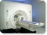
this active shielded member of the Panorama product line combines the advantages of one 1.0 T system's with the possibilities of an open MRI system. The open design helps ease anxiety for claustrophobic patients and increased patient comfort whereby the field strength provides spectacular image quality and fast patient throughput.
Device Information and Specification CLINICAL APPLICATION Whole body Vertically opposed solenoids, head, head-neck, extremity, neck, body/ spine M-XL, shoulder, bilateral breast, wrist, TMJ, flex XS-S-M-L-XL-XXL SE, FE, IR, STIR, FFE, DEFFE, DESE, TSE, DETSE, Single shot SE, DRIVE, Balanced FFE, MRCP, FLAIR, Turbo FLAIR, IR-TSE, T1-STIR TSE, T2-STIR TSE, Diffusion Imaging, 3D SE, 3D FFE, Contrast Perfusion Analysis, MTC;; Angiography: CE-ANGIO, MRA 2D, 3D TOFOpen x 47 cm x infinite (side-first patient entry) POWER REQUIREMENTS 400/480 V | |  | | | |
|  |  | Searchterm 'Spectroscopy' was also found in the following service: | | | | |
|  |  |
| |
|
MRI can be indicated for use in pregnant women if other forms of diagnostic imaging are inadequate or require exposure to ionizing radiation such as X-ray or CT.
As a safety precaution, MR scanning should be avoided in the first three months of pregnancy.
Similar considerations hold for pregnant staff of a magnetic resonance department. An epidemiological study (by Kanal, et al.) concluded that data collected from MRI technologists were negative with respect to any statistically significant elevations in the rates of spontaneous abortion, infertility and premature delivery.
However, also for psychological reasons, it might be a wise precaution that pregnant staff members do not remain in the scan room during actual scanning.
There have been several reports (results could not be reproduced) that static magnetic fields may provoke genetic mutations, changes in growth rate and leukocyte count and other effects.
No reports have been published that persons exposed to magnetic fields, including staff at MR departments, have a higher incidence of genetic damage to their children than found in the average population.
This research needs further investigation and for this purpose pregnancy should be considered a relative contraindication for MR spectroscopy and MRI procedures.
Taking into account that clinical MR imaging devices operate at field strengths of between 0.2 and 2.0 T, higher field strengths need more investigation.

| | | |  | |
• View the DATABASE results for 'Pregnancy' (5).
| | |
• View the NEWS results for 'Pregnancy' (1).
| | | | |  Further Reading: Further Reading: | News & More:
|
|
| |
|  | |  |  |  |
| |
|
A pulse is a rapid change in the amplitude of a RF signal or in some characteristic a RF signal, e.g., phase or frequency, from a baseline value to a higher or lower value, followed by a rapid return to the baseline value. For radio frequencies near the Larmor frequency, it will result in rotation of the macroscopic magnetization vector. The amount of rotation will depend on the strength and duration of the RF pulse; commonly used examples are 90° ( p/2) and 180° ( p) pulses.
RF pulses are used in the spin preparation phase of a pulse sequence, which prepare the spin system for the ensuing measurements. In many sequences, RF pulses are also applied to the volumes outside the one to be measured. This is the case when spatial presaturation techniques are used to suppress artifacts. Many preparation pulses are required in MR spectroscopy to suppress signal from unwanted spins. The simplest preparation pulse making use of spectroscopic properties is a fat saturation pulse, which specifically irradiates the patient at the fat resonant frequency, so that the magnetization coming from fat protons is tilted into the xy-plane where it is subsequently destroyed by a strong dephasing gradient.
The frequency spectrum of RF pulses is critical as it determines the spatial extension and homogeneity over which the spin magnetization is influenced while a gradient field is applied. | |  | |
• View the DATABASE results for 'Radio Frequency Pulse' (16).
| | | | |  Further Reading: Further Reading: | News & More:
|
|
| |
|  |  | Searchterm 'Spectroscopy' was also found in the following services: | | | | |
|  |  |
| |
|
Standard compound used as a standard reference spectral line for defining chemical shifts for a given nucleus. As recommended by the ASTM, for 1H it is tetramethylsilane (TMS) and for 31P it is phosphoric acid, although for practical biological applications water and PCr have been used as secondary references for hydrogen and phosphorus spectroscopy, respectively. The reference compound can be in a capsule outside of the subject (external) or can be in the subject (internal); internal references are generally preferable where possible, as external references may be subject to different conditions. | |  | | | |
|  |  | Searchterm 'Spectroscopy' was also found in the following service: | | | | |
|  |  |
| |
|

(Signa VH/i 3.0T)
With GE Healthcare
leading-edge technology in ultra-high-field imaging. The 3 T VH/i provides a platform for advanced applications in radiology, cardiology, psychology and psychiatry. Real-time image processing lets you acquire multislice whole brain images and map brain functions for research or surgical planning. And the 3 T Signa VH/i is flexible enough to provide clinicians with high performance they require. It can provide not only outstanding features in brain scanning and neuro-system research, but also a wide range of use in scanning breasts, extremities, the spine and the cardiovascular systems.
Device Information and Specification CLINICAL APPLICATION Whole body
T/R quadrature head, T/R quadrature body, T/R phased array extremity (opt) SE, IR, 2D/3D GRE, FGRE, RF-spoiled GRE, FSE, Angiography: 2D/3D TOF, 2D/3D phase contrast vascular IMAGING MODES Single, multislice, volume study, fast scan, multi slab, cine, localizer 100 Images/sec with Reflex100 MULTISLICE 100 Images/sec with Reflex100 2D 0.5-100mm in 0.1mm incremental 128x512 steps 32 phase encode H*W*D 260cm x 238cm x 265cm POWER REQUIREMENTS 480 or 380/415, 3 phase ||
COOLING SYSTEM TYPE Closed-loop water-cooled grad. Less than 0.14 L/hr liquid He | |  | |
• View the DATABASE results for 'Signa 3.0T™' (2).
| | | | |
|  | |  |  |
|  | |
|  | | |
|
| |
 | Look
Ups |
| |