 | Info
Sheets |
| | | | | | | | | | | | | | | | | | | | | | | | |
 | Out-
side |
| | | | |
|
| | | | |
Result : Searchterm 'Suppression' found in 7 terms [ ] and 42 definitions [ ] and 42 definitions [ ] ]
| previous 26 - 30 (of 49) nextResult Pages :  [1 2] [1 2]  [3 4 5 6 7 8 9 10] [3 4 5 6 7 8 9 10] |  | |  | Searchterm 'Suppression' was also found in the following services: | | | | |
|  |  |
| |
|
(DWIBS) This diffusion weighted whole body imaging with background signal suppression provides diffusion weighted contrast throughout the body using a single or multi-station background-suppressed diffusion imaging. DWIBS images are comparable with PET images and provide applications from the visualization of nerve roots and brachial plexus to the detection of lesions throughout the body. See also Diffusion Weighted Imaging (DWI). | |  | | | |  Further Reading: Further Reading: | | Basics:
|
|
News & More:
| |
| |
|  |  | Searchterm 'Suppression' was also found in the following services: | | | | |
|  |  |
| |
|
(DE FGRE, Dual/FFE, DE FFE) Simultaneously acquired in and out of phase TE gradient echo images. To quantitatively measure the signal intensity differences between out of phase and in phase images the parameters should be the same except for the TE.
The chemical shift artifact appearing on the out-of-phase image allows for the detection of lipids in the liver or adrenal gland, such as diffuse fatty infiltration, focal fatty infiltration, focal fatty sparing, benign adrenocortical masses and intracellular lipids within a hepatocellar neoplasm, where spin echo and fat suppression techniques are not as sensitive. Specific pathologies that have been reported include liver lipoma, angiomyolipoma, myelolipoma, metastatic liposarcoma, teratocarcinoma, melanoma, haemorrhagic neoplasm and metastatic choriocarcinoma. | | | |  | |
• View the DATABASE results for 'Dual Echo Fast Gradient Echo' (2).
| | | | |  Further Reading: Further Reading: | News & More:
|
|
| |
|  | |  |  |  |
| |
|

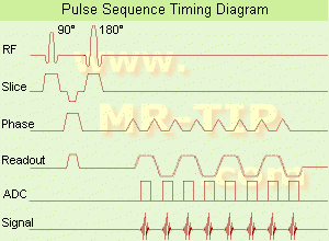
(EPI) Echo planar imaging is one of the early magnetic resonance imaging sequences (also known as Intascan), used in applications like diffusion, perfusion, and functional magnetic resonance imaging. Other sequences acquire one k-space line at each phase encoding step. When the echo planar imaging acquisition strategy is used, the complete image is formed from a single data sample (all k-space lines are measured in one repetition time) of a gradient echo or spin echo sequence (see single shot technique) with an acquisition time of about 20 to 100 ms.
The pulse sequence timing diagram illustrates an echo planar imaging sequence from spin echo type with eight echo train pulses. (See also Pulse Sequence Timing Diagram, for a description of the components.)
In case of a gradient echo based EPI sequence the initial part is very similar to a standard gradient echo sequence. By periodically fast reversing the readout or frequency encoding gradient, a train of echoes is generated.
EPI requires higher performance from the MRI scanner like much larger gradient amplitudes. The scan time is dependent on the spatial resolution required, the strength of the applied gradient fields and the time the machine needs to ramp the gradients.
In EPI, there is water fat shift in the phase encoding direction due to phase accumulations. To minimize water fat shift (WFS) in the phase direction fat suppression and a wide bandwidth (BW) are selected. On a typical EPI sequence, there is virtually no time at all for the flat top of the gradient waveform. The problem is solved by "ramp sampling" through most of the rise and fall time to improve image resolution.
The benefits of the fast imaging time are not without cost. EPI is relatively demanding on the scanner hardware, in particular on gradient strengths, gradient switching times, and receiver bandwidth. In addition, EPI is extremely sensitive to image artifacts and distortions. | |  | |
• View the DATABASE results for 'Echo Planar Imaging' (19).
| | |
• View the NEWS results for 'Echo Planar Imaging' (1).
| | | | |  Further Reading: Further Reading: | Basics:
|
|
| |
|  |  | Searchterm 'Suppression' was also found in the following services: | | | | |
|  |  |
| |
|
(FAT SAT) A specialized technique that selectively saturates fat protons prior to acquiring data as in standard sequences, so that they produce a negligible signal. The presaturation pulse is applied prior to each slice selection. This technique requires a very homogeneous magnetic field and very precise frequency calibration.
Fat saturation does not work well on inhomogeneous volumes of tissue due to a change in the precessional frequencies as the difference in volume affects the magnetic field homogeneity. The addition of a water bag simulates a more homogeneous volume of tissue, thus improving the fat saturation. Since the protons in the water bag are in motion due to recent motion of the bag, phase ghosts can be visualized.
Fat saturation can also be difficult in a region of metallic prosthesis. This is caused by an alteration in the local magnetic field resulting in a change to the precessional frequencies, rendering the chemical saturation pulses ineffective.
See also Fat Suppression, and Dixon. | | | |  | |
• View the DATABASE results for 'Fat Saturation' (9).
| | | | |  Further Reading: Further Reading: | Basics:
|
|
News & More:
| |
| |
|  |  | Searchterm 'Suppression' was also found in the following services: | | | | |
|  |  |
| |
|
(FOV) Defined as the size of the two or three dimensional spatial encoding area of the image. Usually defined in units of mm². The FOV is the square image area that contains the object of interest to be measured. The smaller the FOV, the higher the resolution and the smaller the voxel size but the lower the measured signal.
Useful for decreasing the scantime is a field of view different in the frequency and phase encoding directions ( rectangular field of view - RFOV).
The magnetic field homogeneity decreases as more tissue is imaged (greater FOV). As a result the precessional frequencies change across the imaging volume. That can be a problem for fat suppression imaging. This fat is precessing at the expected frequency only in the center of the imaging volume. E.g. frequency specific fat saturation pulses become less effective when the field of view is increased. It is best to use smaller field of views when applying fat saturation pulses.

Image Guidance
Smaller FOV required higher gradient strength and concludes low signal. Therefore you have to find a compromise between these factors.
The right choice of the field of view is important for MR image quality. When utilizing small field of views and scanning at a distance from the isocenter (more problems with artifacts) it is obviously important to ensure that the region of interest is within the scanning volume.
A smaller FOV in one direction is available with the function rectangular field of view (RFOV).
See also Field Inhomogeneity Artifact. | | | |  | |
• View the DATABASE results for 'Field of View' (27).
| | | | |  Further Reading: Further Reading: | Basics:
|
|
News & More:
| |
| |
|  | |  |  |
|  | | |
|
| |
 | Look
Ups |
| |