Magnetic Resonance - Technology Information Portal Welcome to MRI Technology
Info
  Sheets

Out-
      side


 'T1 Relaxation' 
SEARCH FOR    
 
  2 3 5 A B C D E F G H I J K L M N O P Q R S T U V W X Y Z
Result : Searchterm 'T1 Relaxation' found in 1 term [] and 23 definitions []
previous     21 - 24 (of 24)     
Result Pages : [1]  [2 3 4 5]
Searchterm 'T1 Relaxation' was also found in the following services: 
spacer
News  (2)  Forum  (1)  
 
Spin Echo SequenceInfoSheet: - Sequences - 
Intro, 
Overview, 
Types of, 
etc.MRI Resource Directory:
 - Sequences -
 
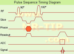
Spin Echo Timing Diagram (SE) The most common pulse sequence used in MR imaging is based of the detection of a spin or Hahn echo. It uses 90° radio frequency pulses to excite the magnetization and one or more 180° pulses to refocus the spins to generate signal echoes named spin echoes (SE).
In the pulse sequence timing diagram, the simplest form of a spin echo sequence is illustrated.
The 90° excitation pulse rotates the longitudinal magnetization (Mz) into the xy-plane and the dephasing of the transverse magnetization (Mxy) starts.
The following application of a 180° refocusing pulse (rotates the magnetization in the x-plane) generates signal echoes. The purpose of the 180° pulse is to rephase the spins, causing them to regain coherence and thereby to recover transverse magnetization, producing a spin echo.
The recovery of the z-magnetization occurs with the T1 relaxation time and typically at a much slower rate than the T2-decay, because in general T1 is greater than T2 for living tissues and is in the range of 100-2000 ms.
The SE pulse sequence was devised in the early days of NMR days by Carr and Purcell and exists now in many forms: the multi echo pulse sequence using single or multislice acquisition, the fast spin echo (FSE/TSE) pulse sequence, echo planar imaging (EPI) pulse sequence and the gradient and spin echo (GRASE) pulse sequence;; all are basically spin echo sequences.
In the simplest form of SE imaging, the pulse sequence has to be repeated as many times as the image has lines.
Contrast values:
PD weighted: Short TE (20 ms) and long TR.
T1 weighted: Short TE (10-20 ms) and short TR (300-600 ms)
T2 weighted: Long TE (greater than 60 ms) and long TR (greater than 1600 ms)
With spin echo imaging no T2* occurs, caused by the 180° refocusing pulse. For this reason, spin echo sequences are more robust against e.g., susceptibility artifacts than gradient echo sequences.

See also Pulse Sequence Timing Diagram to find a description of the components.
 
Images, Movies, Sliders:
 Shoulder Coronal T1 SE  Open this link in a new window
    

Courtesy of  Robert R. Edelman
 Shoulder Axial T1 SE  Open this link in a new window
 MRI Orbita T1  Open this link in a new window
    
 
spacer
 
• Related Searches:
    • Echo Time
    • Brain MRI
    • Gradient
    • Repetition Time
    • T2 Star
 
Further Reading:
  Basics:
Fast Spin Echo(.pdf)
Tuesday, 24 January 2006   by www.81bones.net    
Magnetic resonance imaging
   by www.scholarpedia.org    
FUNDAMENTALS OF MRI: Part I
   by www.e-radiography.net    
  News & More:
New MR sequence helps radiologists more accurately evaluate abnormalities of the uterus and ovaries
Thursday, 23 April 2009   by www.eurekalert.org    
MRI techniques improve pulmonary embolism detection
Monday, 19 March 2012   by medicalxpress.com    
MRI Resources 
Musculoskeletal and Joint MRI - Pathology - Services and Supplies - Claustrophobia - Case Studies - Safety Training
 
Stimulated Echo
 
A form of a spin echo produced by three pulse RF sequences, consisting of two RF pulses following an initial exciting RF pulse. The stimulated echo appears at a time delay after the third pulse equal to the interval between the first two pulses. Although classically produced with 90° pulses, any RF pulses other than an ideal 180° can produce a stimulated echo. The intensity of the echo depends in part on the T1 relaxation time because the excitation is 'stored' as longitudinal magnetization between the second and third RF pulses. For example, use of stimulated echoes with spatially selective excitation with orthogonal magnetic field gradients permits volume-selective excitation for spectroscopic localization.
mri safety guidance
Image Guidance
Artifacts may appear as a series of fine lines. A narrow bandwidth causes a wide read window, which allows the stimulated echo to be incorporated into the image data. This can be supported by increasing the received bandwidth, which would narrow the read window, thus not incorporating the extraneous echo. Another help would be to change the first echo time, which may change the spacing of the stimulated echoes to outside that of the read window for the second echo.
spacer

• View the DATABASE results for 'Stimulated Echo' (8).Open this link in a new window

 
Further Reading:
  Basics:
Magnetic resonance imaging
   by www.scholarpedia.org    
Clinical evaluation of a speed optimized T2 weighted fast spin echo sequence at 3.0 T using variable flip angle refocusing, half-Fourier acquisition and parallel imaging
Wednesday, 25 October 2006
MRI Resources 
Safety Products - Pregnancy - Databases - Online Books - Raman Spectroscopy - Shielding
 
Superparamagnetic Contrast AgentsInfoSheet: - Contrast Agents - 
Intro, Overview, 
Characteristics, 
Types of, 
etc.MRI Resource Directory:
 - Contrast Agents -
 
Small particles of ferrite are used as superparamagnetic contrast medium in MR imaging (appearing predominantly dark on MRI). These agents exhibit strong T1 relaxation properties, and due to susceptibility differences to their surroundings also produce a strongly varying local magnetic field, which enhances T2 relaxation to darken the contrast media containing structures.
Superparamagnetic contrast agents are also known by the abbreviation SPIO's (small particle iron oxide or superparamagnetic iron oxide) and USPIO's (ultrasmall particle iron oxide or ultrasmall superparamagnetic iron oxide).
Two types of USPIO will be available on the market as blood pool agents, while SPIO's have been used as darkening contrast agents for liver imaging. As particulate matter they are taken up by the RES. Very small particles of less than 300 nanometers also remain intravascular for a prolonged period of time and thus can serve as blood pool agents.

See also the related poll result: 'The development of contrast agents in MRI is'
spacer

• View the DATABASE results for 'Superparamagnetic Contrast Agents' (12).Open this link in a new window

 
Further Reading:
  News & More:
Optimized Labelling of Human Monocytes with Iron Oxide MR Contrast Agents
Sunday, 30 November 2003   by rsna2003.rsna.org    
Poly (dopamine) coated superparamagnetic iron oxide nanocluster for noninvasive labeling, tracking, and targeted delivery of adipose tissue-derived stem cells
Tuesday, 5 January 2016   by www.nature.com    
Searchterm 'T1 Relaxation' was also found in the following services: 
spacer
News  (2)  Forum  (1)  
 
Teslascan®InfoSheet: - Contrast Agents - 
Intro, Overview, 
Characteristics, 
Types of, 
etc.MRI Resource Directory:
 - Contrast Agents -
 
(Mn-DPDP) This agent, mangafodipir trisodium, is a hepatocyte specific MRI contrast agent. Manganese is very toxic, so it has to be chelated and put in the form of a vitamin B6 analog, which is taken up by normal hepatocytes to some extent.
Teslascan® was developed in the early 1980's, went through clinical trials in the early 1990's, and was approved in 1997. One problem with assessing the efficacy of this agent is the fact that the phase III trials finished in the early 1990's, and the techniques used for MR today are very different from the techniques used almost a decade ago.
This contrast agent shortens the T1 relaxation time. On T1 weighted pictures it makes a normal liver look brighter. Since metastases, for example, do not generally take up this agent, the contrast between the enhancing liver and the non-enhancing lesions will increase on T1 weighted pictures. It does not have much effect on T2 weighted images.
Drug Information and Specification
NAME OF COMPOUND
Mangafodipir trisodium, Manganese dipyroxyl diphosphate, MN-DPDP
DEVELOPER
CENTRAL MOIETY
Mn2+
CONTRAST EFFECT
T1, Predominantly positive enhancement
r1=2.3, r2=4.0, B0=1.0 T
PHARMACOKINETIC
Hepatobiliary, pancreatic, adrenal
290 mosm/kgH2O
CONCENTRATION
0.01 mmol/L
DOSAGE
5 µmol/kg, 0.5 ml/kg
PREPARATION
Finished product
INDICATION
Liver lesions
DEVELOPMENT STAGE
Approved
DISTRIBUTOR
See below
PRESENTATION
Vials of 100 ml
DO NOT RELY ON THE INFORMATION PROVIDED HERE, THEY ARE
NOT A SUBSTITUTE FOR THE ACCOMPANYING PACKAGE INSERT!
Distribution Information
TERRITORY
TRADE NAME
DEVELOPMENT
STAGE
DISTRIBUTOR
USA
Teslascan®
for sale
EU
Teslascan®
for sale
spacer

• View the DATABASE results for 'Teslascan®' (4).Open this link in a new window

 
Further Reading:
  Basics:
EMEA - Teslascan - SCIENTIFIC DISCUSSION(.pdf)
   by www.emea.europa.eu    
  News & More:
Diagnosis and staging of pancreatic cancer: comparison of mangafodipir trisodium-enhanced MR imaging and contrast-enhanced helical hydro-CT.
2002
MAGNETIC RESONANCE IMAGING OF FOCAL LIVER LESIONS(.pdf)
2002
MRI Resources 
Jobs pool - Libraries - Fluorescence - Claustrophobia - IR - Diffusion Weighted Imaging
 
previous      21 - 24 (of 24)     
Result Pages : [1]  [2 3 4 5]
 Random Page
 
Share This Page
FacebookTwitterLinkedIn

MR-TIP    
Community   
User
Pass
Forgot your UserID/Password ?    


MRI is trending to low field magnets :
reduced costs will lead to this change 
AI will close the gap to high field 
only in remote areas 
is only temporary 
never 

Look
      Ups


MR-TIP.com uses cookies! By browsing MR-TIP.com, you agree to our use of cookies.

Magnetic Resonance - Technology Information Portal
Member of SoftWays' Medical Imaging Group - MR-TIP • Radiology-TIP • Medical-Ultrasound-Imaging • 
Copyright © 2003 - 2024 SoftWays. All rights reserved. [ 21 November 2024]
Terms of Use | Privacy Policy | Advertising
 [last update: 2024-02-26 03:41:00]