Magnetic Resonance - Technology Information Portal Welcome to MRI Technology
Info
  Sheets

Out-
      side


 'Transverse Magnetization' 
SEARCH FOR    
 
  2 3 5 A B C D E F G H I J K L M N O P Q R S T U V W X Y Z
Result : Searchterm 'Transverse Magnetization' found in 1 term [] and 42 definitions []
previous     11 - 15 (of 43)     next
Result Pages : [1]  [2 3 4 5 6 7 8 9]
MRI Resources 
Services and Supplies - Contrast Agents - Open Directory Project - Corporations - Health - Nerve Stimulator
 
Spin Echo SequenceInfoSheet: - Sequences - 
Intro, 
Overview, 
Types of, 
etc.MRI Resource Directory:
 - Sequences -
 
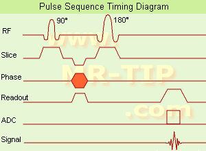
Spin Echo Timing Diagram (SE) The most common pulse sequence used in MR imaging is based of the detection of a spin or Hahn echo. It uses 90° radio frequency pulses to excite the magnetization and one or more 180° pulses to refocus the spins to generate signal echoes named spin echoes (SE).
In the pulse sequence timing diagram, the simplest form of a spin echo sequence is illustrated.
The 90° excitation pulse rotates the longitudinal magnetization (Mz) into the xy-plane and the dephasing of the transverse magnetization (Mxy) starts.
The following application of a 180° refocusing pulse (rotates the magnetization in the x-plane) generates signal echoes. The purpose of the 180° pulse is to rephase the spins, causing them to regain coherence and thereby to recover transverse magnetization, producing a spin echo.
The recovery of the z-magnetization occurs with the T1 relaxation time and typically at a much slower rate than the T2-decay, because in general T1 is greater than T2 for living tissues and is in the range of 100-2000 ms.
The SE pulse sequence was devised in the early days of NMR days by Carr and Purcell and exists now in many forms: the multi echo pulse sequence using single or multislice acquisition, the fast spin echo (FSE/TSE) pulse sequence, echo planar imaging (EPI) pulse sequence and the gradient and spin echo (GRASE) pulse sequence;; all are basically spin echo sequences.
In the simplest form of SE imaging, the pulse sequence has to be repeated as many times as the image has lines.
Contrast values:
PD weighted: Short TE (20 ms) and long TR.
T1 weighted: Short TE (10-20 ms) and short TR (300-600 ms)
T2 weighted: Long TE (greater than 60 ms) and long TR (greater than 1600 ms)
With spin echo imaging no T2* occurs, caused by the 180° refocusing pulse. For this reason, spin echo sequences are more robust against e.g., susceptibility artifacts than gradient echo sequences.

See also Pulse Sequence Timing Diagram to find a description of the components.
 
Images, Movies, Sliders:
 Shoulder Coronal T1 SE  Open this link in a new window
    

Courtesy of  Robert R. Edelman
 Shoulder Axial T1 SE  Open this link in a new window
 MRI Orbita T1  Open this link in a new window
    
 
spacer
 
• Related Searches:
    • T2 Weighted
    • Pulse Sequence Timing Diagram
    • Brain MRI
    • Lumbar Spine MRI
    • T2 Time
 
Further Reading:
  Basics:
Fast Spin Echo(.pdf)
Tuesday, 24 January 2006   by www.81bones.net    
Magnetic resonance imaging
   by www.scholarpedia.org    
FUNDAMENTALS OF MRI: Part I
   by www.e-radiography.net    
  News & More:
New MR sequence helps radiologists more accurately evaluate abnormalities of the uterus and ovaries
Thursday, 23 April 2009   by www.eurekalert.org    
MRI techniques improve pulmonary embolism detection
Monday, 19 March 2012   by medicalxpress.com    
MRI Resources 
Implant and Prosthesis pool - Services and Supplies - Journals - Chemistry - MRCP - Universities
 
T1 Weighted ImageInfoSheet: - Sequences - 
Intro, 
Overview, 
Types of, 
etc.
 
The basis of T1 weighted imaging is the longitudinal relaxation. A T1 weighted magnetic resonance image is created typically by using short TE and TR times.
The final image is a reflection of more than one of these pulse sequence parameters, weighted according to the type of sequence and its timing. T1 signals determine predominantly the contrast and brightness in this type of images but proton density will always contribute to the image intensity. The T1 dependence is mainly determined by the repetition time or any pre-pulses (such as in an inversion recovery pulse sequence).
Due to the larger longitudinal and transverse magnetization, fat has a higher signal and will appear bright on a T1 contrast MR image. Conversely, water has less longitudinal magnetization prior to a RF pulse, therefore less transverse magnetization after a RF pulse yielding low signal appearing dark on a T1 contrast image. Often, a paramagnetic contrast agent, a gadolinium compound, is administered, and both pre-contrast T1 weighted images and post-contrast T1 weighted images are obtained.
 
Images, Movies, Sliders:
 Lumbar Spine T1 SE Sagittal  Open this link in a new window
    

Courtesy of  Robert R. Edelman
 Shoulder Axial T1 SE  Open this link in a new window
 Shoulder Coronal T1 SE  Open this link in a new window
    

Courtesy of  Robert R. Edelman
 
spacer

• View the DATABASE results for 'T1 Weighted Image' (5).Open this link in a new window

 
Further Reading:
  Basics:
Magnetic resonance imaging - From Wikipedia, the free encyclopedia.
   by en.wikipedia.org    
  News & More:
Possible New MRI Marker for Multiple Sclerosis Progression
Wednesday, 29 August 2007   by www.medpagetoday.com    
MRI Resources 
Pediatric and Fetal MRI - Education pool - Societies - Intraoperative MRI - Education - Stimulator pool
 
T2 TimeForum -
related threads
 
The T2 relaxation time (spin spin relaxation time or transverse relaxation time), is a biological parameter that is used in MRIs to distinguish between tissue types and is termed 'Time 2' or T2. It is a tissue-specific time constant for protons and is dependent on the exchanging of energy with near by nuclei. T2 weighted images rely upon local dephasing of spins following the application of the transverse energy pulse. T2 is the decay of magnetization perpendicular to the main magnetic field (in an ideal homogeneous field).
Due to interaction between the spins, they lose their phase coherence, which results in a loss of transverse magnetization and MRI signal. After time T2 transverse magnetization has lost 63% of its original value. This tissue parameter determines the contrast.
The T2 relaxation is temperature dependent. At a lower temperature molecular motion is reduced and the decay times are reduced.
Fat has a very efficient energy exchange and therefore it has a relatively short T2.
Water is less efficient than fat in the exchange of energy, and therefore it has a long T2 time.

See also T2 Weighted Image and Magnetic Resonance Imaging MRI.
 
Images, Movies, Sliders:
 Breast MRI Images T2 And T1  Open this link in a new window
      
 MRI of the Skull Base  Open this link in a new window
    
SlidersSliders Overview

 Knee MRI Transverse 002  Open this link in a new window
    
 
spacer

• View the DATABASE results for 'T2 Time' (16).Open this link in a new window

 
Further Reading:
  News & More:
MYELIN-SELECTIVE MRI: PULSE SEQUENCE DESIGN AND OPTIMIZATION
   by www.imaging.robarts.ca    
MRI Resources 
Developers - Stent - Breast MRI - Artifacts - Collections - Directories
 
Transverse Relaxation Time
 
The T2 time constant, which determines the rate at which excited protons reach equilibrium, or go out of phase with each other. A measure of the time taken for spinning protons to lose phase coherence among the nuclei spinning perpendicular to the main field due to interaction between spins, resulting in a reduction in the transverse magnetization. The transverse magnetization value will drop from maximum to a value of about 37% of its original value in a time of T2.
 
Images, Movies, Sliders:
 Brain MRI Images Axial T2  Open this link in a new window
      

 Breast MRI 2 Transverse T2 001  Open this link in a new window
 
spacer

• View the DATABASE results for 'Transverse Relaxation Time' (4).Open this link in a new window

MRI Resources 
MRI Reimbursement - Artifacts - Health - Jobs - Spine MRI - Process Analysis
 
Balanced Fast Field EchoInfoSheet: - Sequences - 
Intro, 
Overview, 
Types of, 
etc.MRI Resource Directory:
 - Sequences -
 
(bFFE) A FFE sequence using a balanced gradient waveform. A balanced sequence starts out with a RF pulse of 90° or less and the spins in the steady state. Before the next TR in the slice phase and frequency encoding, gradients are balanced so their net value is zero. Now the spins are prepared to accept the next RF pulse, and their corresponding signal can become part of the new transverse magnetization. Since the balanced gradients maintain the transverse and longitudinal magnetization, the result is, that both T1 and T2 contrast are represented in the image. This pulse sequence produces images with increased signal from fluid, along with retaining T1 weighted tissue contrast. Because this form of sequence is extremely dependent on field homogeneity, it is essential to run a shimming prior the acquisition. A fully balanced (refocused) sequence would yield higher signal, especially for tissues with long T2 relaxation times.

See Steady State Free Precession and Gradient Echo Sequence.
 
Images, Movies, Sliders:
 Cardiac Infarct Short Axis Cine bFFE 1  Open this link in a new window
    
 
spacer

• View the DATABASE results for 'Balanced Fast Field Echo' (3).Open this link in a new window

 
Further Reading:
  News & More:
T1rho-prepared balanced gradient echo for rapid 3D T1rho MRI
Monday, 1 September 2008   by www.ncbi.nlm.nih.gov    
Utility of the FIESTA Pulse Sequence in Body Oncologic Imaging: Review
June 2009   by www.ajronline.org    
MRI Resources 
Quality Advice - MRI Reimbursement - Diffusion Weighted Imaging - Bioinformatics - Manufacturers - Abdominal Imaging
 
previous      11 - 15 (of 43)     next
Result Pages : [1]  [2 3 4 5 6 7 8 9]
 Random Page
 
Share This Page
FacebookTwitterLinkedIn

MR-TIP    
Community   
User
Pass
Forgot your UserID/Password ?    


Personalized protocols (age, gender, body habitus, etc.) lead to :
more automated planning 
improved patient comfort 
better diagnostics 
optimized image quality 
nothing 

Look
      Ups


MR-TIP.com uses cookies! By browsing MR-TIP.com, you agree to our use of cookies.

Magnetic Resonance - Technology Information Portal
Member of SoftWays' Medical Imaging Group - MR-TIP • Radiology-TIP • Medical-Ultrasound-Imaging • 
Copyright © 2003 - 2024 SoftWays. All rights reserved. [ 21 November 2024]
Terms of Use | Privacy Policy | Advertising
 [last update: 2024-02-26 03:41:00]