Magnetic Resonance - Technology Information Portal Welcome to MRI Technology
Info
  Sheets

Out-
      side


 'Trigger' 
SEARCH FOR    
 
  2 3 5 A B C D E F G H I J K L M N O P Q R S T U V W X Y Z
Result : Searchterm 'Trigger' found in 6 terms [] and 15 definitions []
previous     16 - 20 (of 21)     next
Result Pages : [1 2]  [3 4 5]
Searchterm 'Trigger' was also found in the following services: 
spacer
News  (3)  Forum  (3)  
 
Audio Frequency ArtifactInfoSheet: - Artifacts - 
Case Studies, 
Reduction Index, 
etc.MRI Resource Directory:
 - Artifacts -
 
Quick Overview
Artifact Information
NAME
Audio frequency
DESCRIPTION
Ghosting, lines or spots
REASON
Wrong modulation at audio rate, wrong audio signal
HELP
AC-line synchronization
Two types of audio-frequency problems are possible:
1. Modulation of the MR signal at an audio rate
2. Audio signal component at digitizer input
Problem 1 looks like ghosts, weak copies of the real image, displaced along the phase encoding direction. The number and intensity of the ghosts depends upon the relationship between the period of the audio modulation and the repetition time.
Problem 2 shows up as lines or spots at the appropriate points along the frequency direction. If there is no correlation between the audio period and TR, lines are generated or discrete spots occur.
mri safety guidance
Image Guidance
Both problems can be lessened by use of AC-line synchronization (line trigger).
spacer
Searchterm 'Trigger' was also found in the following services: 
spacer
Radiology  (5) Open this link in a new windowUltrasound  (4) Open this link in a new window
Double Inversion Recovery T1 MeasurementInfoSheet: - Sequences - 
Intro, 
Overview, 
Types of, 
etc.MRI Resource Directory:
 - Sequences -
 
(DIR or DIRT1) Double inversion recovery T1 measurement is a T1 weighted black blood MRA sequence in which the signal from blood is suppressed. The inversion time to suppress blood is described as the duration between the initial inversion pulse and time point that the longitudinal magnetization of blood reaches the zero point. The readout starts at the blood suppression inversion time (BSP TI) and blood in the imaging slice gives no signal. This inversion time is around 650 ms with a 60 beat per minute heart rate at 1.5 T.
The TI can be decreased by using a wider receive bandwidth, shorter echo train length and/or narrow trigger window. Wide bandwidth also decreases the blurring caused by long echo trains at the expense of signal to noise ratio. In case of in plane or slow flow the suppression of the signal from blood may be incomplete. With increased TE or change of the image plane the blood suppression can be improved.
Double inversion recovery is a breath hold technique with one image per acquisition used in cardiovascular imaging. The patient is instructed to hold the breath in expiration (if not possible also inspiration can be taken), so that the end diastolic volume in the cardiac chambers would be the same during entire scanning. DIR provides fine details of the boundary between the lumen and the wall of the cardiac chambers and main vascular and heart structures, pericardium, and mediastinal tissues.
 
Images, Movies, Sliders:
 Normal Dual Inversion Fast Spin-echo  Open this link in a new window
      

Courtesy of  Robert R. Edelman

 
spacer

• View the DATABASE results for 'Double Inversion Recovery T1 Measurement' (2).Open this link in a new window

 
Further Reading:
  News & More:
Artificial double inversion recovery images can substitute conventionally acquired images: an MRI-histology study
Wednesday, 16 February 2022   by www.nature.com    
MRI Resources 
Homepages - Corporations - Distributors - Contrast Agents - Most Wanted - Services and Supplies
 
Echelon™ 1.5TInfoSheet: - Devices -
Intro, 
Types of Magnets, 
Overview, 
etc.
 
www.hitachimed.com/contentindex.asp?ID=971 From Hitachi Medical Systems America Inc.;
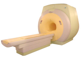
Hitachi expanded its portfolio with the Echelon™ 1.5T. The MRI scanner combines a compact magnet and a scalable 8-channel RF system with high-performance gradients and slew rate to select short echo times, small field of views, high matrices and thin slices. Standard features of the Echelon MRI system include higher-order active shim, RAPID (parallel imaging for use on brain MRI, body, cardiovascular imaging, and orthopedic coils), multiple coil ports, and an advanced reconstruction engine.
Device Information and Specification
CLINICAL APPLICATION
Whole body
CONFIGURATION
Short bore
Head, body coil, spine, breast, knee, shoulder, vascular multiple array coils.
SYNCHRONIZATION
Cardiac gating, ECG/peripheral, respiratory gating
PULSE SEQUENCES
SE, IR, FSE, FIR, GE, SG, BASG, PBSG, PCIR, DWI, Radial, Angiography: TOF, FLUTE (Fluoro-triggered bolus MRA), Time-resolved MRA
IMAGING MODES
Single, multislice, volume study
PIXEL INTENSITY
Level Range: -2,000 to +4,000
Sub millimeter
POWER REQUIREMENTS
208/220/240 V, single phase
CRYOGEN USE
Low cryogen boil-off
STRENGTH
30 mT/m
150 T/m/sec
Higher-order active shim
spacer

• View the DATABASE results for 'Echelon™ 1.5T' (2).Open this link in a new window


• View the NEWS results for 'Echelon™ 1.5T' (3).Open this link in a new window.
 
Further Reading:
  Basics:
Echelon 1.5T
   by www.hitachimed.com    
Searchterm 'Trigger' was also found in the following services: 
spacer
News  (3)  Forum  (3)  
 
FORTE 3.0T™InfoSheet: - Devices -
Intro, 
Types of Magnets, 
Overview, 
etc.MRI Resource Directory:
 - Devices -
 
www.isoltech.co.kr/english/product/30t.htm From ISOL Technology
'Ultra high field MR system, it's right close to you. FORTE 3.0T is the new standard for the future ultra high field MR system. If you are pushing the limits of your existing clinical MR scanner, the FORTE will surely take you to the next level of diagnostic imaging. FORTE is the core leader of the medical technology in the 21st century. Proving effects of fMRI that cannot be measured with MRI less than 2.0T.'
Device Information and Specification
CLINICAL APPLICATION
Whole body
CONFIGURATION
Short bore compact
SYNCHRONIZATION
ECG/peripheral: Optional/yes, external trigger, respiratory gating
PULSE SEQUENCES
Spin echo, Gradient echo, Fast spin echo, Inversion recovery, 2D/3D Fast gradient echo sequences FLAIR/STIR, 2D/3D TOF
IMAGING MODES
2D/3D, T1, T2 and Diffusion//Perfusion imaging, MR Angiography package, Advanced EPI package, Multi-nuclei MR Spectroscopy package
FOV
40 cm
128 x 128, 256 x 256, 512 x 512, 1024 x 1024
BORE DIAMETER
or W x H
61 cm without body coil
MAGNET WEIGHT
12000 kg
H*W*D
260 x 220 x 235 cm
COOLING SYSTEM TYPE
Water-cooled coil and air-cooled amplifier
CRYOGEN USE
0.15 L/hr helium
STRENGTH
38 mT/m
5-GAUSS FRINGE FIELD
3.3 m / 5.2 m
Passive and active
spacer

• View the DATABASE results for 'FORTE 3.0T™' (2).Open this link in a new window

Searchterm 'Trigger' was also found in the following services: 
spacer
Radiology  (5) Open this link in a new windowUltrasound  (4) Open this link in a new window
Lung ImagingMRI Resource Directory:
 - Lung Imaging -
 
Lung imaging is furthermore a challenge in MRI because of the predominance of air within the lungs and associated susceptibility issues as well as low signal to noise of the inflated lung parenchyma. Cardiac and respiratory triggered or breath hold sequences allow diagnostic imaging, however a comparable image quality with computed tomography is still difficult to achieve.
Assumptions for lung MRI:
•
Low signal to noise ratio of the inherently low lung proton density.
•
Cardiac and respiratory motion artifacts.
•
Magnetic susceptibility effects of large magnetic field gradients.
•
Very short transverse relaxation times and significant diffusion yielding short T2 (30-70 msec), short T2* (1-3 msec), and additional long T1 relaxation times (1300-1500 msec).
•
The extreme short T2 values are responsible for a fast signal decay during a single shot readout, resulting in blurring.

The current trends in MRI are the use of new imaging technologies and increasingly powerful magnetic fields. Among these technologies are parallel imaging techniques as well as ventilation agents like hyperpolarized helium for the use as an inert inhalational contrast agent to study lung ventilation properties. With hyperpolarized gases clear images of the lungs can be obtained without using a large magnetic field (see also back projection imaging). Single shot sequences (e.g. TSE or Half Fourier Acquisition Single Shot Turbo Spin Echo HASTE) used in lung MR imaging benefits from parallel imaging techniques due to reduced relaxation time effects during the echo train and therefore reduced image blurring as well as reduced motion artifacts.
In the future, more effective contrast agents may provide an alternative solution to the need for high field MRI. Dynamic contrast enhanced MRI perfusion has demonstrated a potential in the diagnosis of pulmonary embolism or to characterize lung cancer and mediastinal tumors. 3D contrast enhanced magnetic resonance angiography of the thoracic vessel.

See also the related poll result: 'MRI will have replaced 50% of x-ray exams by'
 
Images, Movies, Sliders:
 Anatomic Imaging of the Lungs  Open this link in a new window
      

Courtesy of  Robert R. Edelman
 Normal Lung Gd Perfusion MRI  Open this link in a new window
      

Courtesy of  Robert R. Edelman

 MRI Thorax Basal Plane  Open this link in a new window
 
Radiology-tip.comradLung Scintigraphy
spacer

• View the DATABASE results for 'Lung Imaging' (7).Open this link in a new window


• View the NEWS results for 'Lung Imaging' (3).Open this link in a new window.
 
Further Reading:
  Basics:
A safer approach for diagnostic medical imaging
Monday, 29 September 2014   by www.eurekalert.org    
Parallel Lung Imaging(.pdf)
  News & More:
Chest MRI a viable alternative to chest CT in COVID-19 pneumonia follow-up
Monday, 21 September 2020   by www.healthimaging.com    
CT Imaging Features of 2019 Novel Corona virus (2019-nCoV)
Tuesday, 4 February 2020   by pubs.rsna.org    
Polarean Imaging Phase III Trial Results Point to Potential Improvements in Lung Imaging
Wednesday, 29 January 2020   by www.diagnosticimaging.com    
Low Power MRI Helps Image Lungs, Brings Costs Down
Thursday, 10 October 2019   by www.medgadget.com    
Chest MRI Using Multivane-XD, a Novel T2-Weighted Free Breathing MR Sequence
Thursday, 11 July 2019   by www.sciencedirect.co    
Researchers Review Importance of Non-Invasive Imaging in Diagnosis and Management of PAH
Wednesday, 11 March 2015   by lungdiseasenews.com    
New MRI Approach Reveals Bronchiectasis' Key Features Within the Lung
Thursday, 13 November 2014   by lungdiseasenews.com    
MRI techniques improve pulmonary embolism detection
Monday, 19 March 2012   by medicalxpress.com    
  News & More:
Partnership with VIDA to streamline adoption of advanced MRI of the lungs
Monday, 11 September 2023   by www.itnonline.com    
MRI Resources 
Knee MRI - Chemistry - Education pool - MR Guided Interventions - Coils - Absorption and Emission
 
previous      16 - 20 (of 21)     next
Result Pages : [1 2]  [3 4 5]
 Random Page
 
Share This Page
FacebookTwitterLinkedIn

MR-TIP    
Community   
User
Pass
Forgot your UserID/Password ?    


New acceleration techniques will :
reduce scan times 
cause artifacts 
increase expenses 
be useful if you have a lot of experience 
doesn't do much 
never heard of 

Look
      Ups


MR-TIP.com uses cookies! By browsing MR-TIP.com, you agree to our use of cookies.

Magnetic Resonance - Technology Information Portal
Member of SoftWays' Medical Imaging Group - MR-TIP • Radiology-TIP • Medical-Ultrasound-Imaging • 
Copyright © 2003 - 2024 SoftWays. All rights reserved. [ 21 November 2024]
Terms of Use | Privacy Policy | Advertising
 [last update: 2024-02-26 03:41:00]