Magnetic Resonance - Technology Information Portal Welcome to MRI Technology
Info
  Sheets

Out-
      side


 'pulse sequence' 
SEARCH FOR    
 
  2 3 5 A B C D E F G H I J K L M N O P Q R S T U V W X Y Z
Result : Searchterm 'pulse sequence' found in 5 terms [] and 166 definitions []
previous     21 - 25 (of 171)     next
Result Pages : [1]  [2 3 4 5 6 7 8 9 10 11 12 13 14 15 16 17 18 19 20 ... ]
Searchterm 'pulse sequence' was also found in the following services: 
spacer
News  (2)  Resources  (5)  Forum  (10)  
 
ContrastForum -
related threads
 
Contrast is the relative difference of signal intensities in two adjacent regions of an image.
Due to the T1 and T2 relaxation properties in magnetic resonance imaging, differentiation between various tissues in the body is possible. Tissue contrast is affected by not only the T1 and T2 values of specific tissues, but also the differences in the magnetic field strength, temperature changes, and many other factors. Good tissue contrast relies on optimal selection of appropriate pulse sequences (spin echo, inversion recovery, gradient echo, turbo sequences and slice profile).
Important pulse sequence parameters are TR (repetition time), TE (time to echo or echo time), TI (time for inversion or inversion time) and flip angle. They are associated with such parameters as proton density and T1 or T2 relaxation times. The values of these parameters are influenced differently by different tissues and by healthy and diseased sections of the same tissue.
For the T1 weighting it is important to select a correct TR or TI. T2 weighted images depend on a correct choice of the TE. Tissues vary in their T1 and T2 times, which are manipulated in MRI by selection of TR, TI, and TE, respectively. Flip angles mainly affect the strength of the signal measured, but also affect the TR/TI/TE parameters.
Conditions necessary to produce different weighted images:
T1 Weighted Image: TR value equal or less than the tissue specific T1 time - TE value less than the tissue specific T2 time.
T2 Weighted Image: TR value much greater than the tissue specific T1 time - TE value greater or equal than the tissue specific T2 time.
Proton Density Weighted Image: TR value much greater than the tissue specific T1 time - TE value less than the tissue specific T2 time.

See also Image Contrast Characteristics, Contrast Reversal, Contrast Resolution, and Contrast to Noise Ratio.
 
Images, Movies, Sliders:
 Fetus (Brain) and Dermoid in Mother  Open this link in a new window
      

Courtesy of  Robert R. Edelman

 Circle of Willis, Time of Flight, MIP  Open this link in a new window
    
SlidersSliders Overview

 Anatomic MRI of the Knee 1  Open this link in a new window
    
SlidersSliders Overview

 Anatomic Imaging of the Liver  Open this link in a new window
      

 Brain MRI Inversion Recovery  Open this link in a new window
    
 
spacer
 
• Related Searches:
    • Contrast Medium
    • Repetition Time
    • Contrast to Noise Ratio
    • Proton Density
    • Medical Imaging
 
Further Reading:
  Basics:
Magnetic resonance imaging
   by www.scholarpedia.org    
MRI's inside story
Thursday, 4 December 2003   by www.economist.com    
Image Characteristics and Quality
   by www.sprawls.org    
  News & More:
A natural boost for MRI scans
Monday, 21 October 2013   by www.eurekalert.org    
A groundbreaking new graphene-based MRI contrast agent
Friday, 8 June 2012   by www.nanowerk.com    
New MRI Chemical Offers Amazing Contrast
Friday, 22 January 2010   by news.softpedia.com    
Searchterm 'pulse sequence' was also found in the following service: 
spacer
Ultrasound  (1) Open this link in a new window
Diffusion Weighted ImagingForum -
related threadsMRI Resource Directory:
 - Diffusion Weighted Imaging -
 
(DWI) Magnetic resonance imaging is sensitive to diffusion, because the diffusion of water molecules along a field gradient reduces the MR signal. In areas of lower diffusion the signal loss is less intense and the display from this areas is brighter. The use of a bipolar gradient pulse and suitable pulse sequences permits the acquisition of diffusion weighted images (images in which areas of rapid proton diffusion can be distinguished from areas with slow diffusion).
Based on echo planar imaging, multislice DWI is today a standard for imaging brain infarction. With enhanced gradients, the whole brain can be scanned within seconds. The degree of diffusion weighting correlates with the strength of the diffusion gradients, characterized by the b-value, which is a function of the gradient related parameters: strength, duration, and the period between diffusion gradients.
Certain illnesses show restrictions of diffusion, for example demyelinization and cytotoxic edema. Areas of cerebral infarction have decreased apparent diffusion, which results in increased signal intensity on diffusion weighted MRI scans. DWI has been demonstrated to be more sensitive for the early detection of stroke than standard pulse sequences and is closely related to temperature mapping.
DWIBS is a new diffusion weighted imaging technique for the whole body that produces PET-like images. The DWIBS sequence has been developed with the aim to detect lymph nodes and to differentiate normal and hyperplastic from metastatic lymph nodes. This may be possible caused by alterations in microcirculation and water diffusivity within cancer metastases in lymph nodes.

See also Diffusion Weighted Sequence, Perfusion Imaging, ADC Map, Apparent Diffusion Coefficient, and Diffusion Tensor Imaging.
spacer

• View the DATABASE results for 'Diffusion Weighted Imaging' (11).Open this link in a new window


• View the NEWS results for 'Diffusion Weighted Imaging' (4).Open this link in a new window.
 
Further Reading:
  Basics:
EVALUATION OF HUMAN STROKE BY MR IMAGING
2000
Novel MRI Technique Could Reduce Breast Biopsies, University of Washington Study
Tuesday, 2 October 2012   by www.eurekalert.org    
Quantitative Apparent Diffusion Coefficient Measurements Obtained by 3-Tesla MRI Are Correlated with Biomarkers of Bladder Cancer Proliferative Activity
   by www.plosone.org    
  News & More:
Stability and repeatability of diffusion-weighted imaging (DWI) of normal pancreas on 5.0 Tesla magnetic resonance imaging (MRI)
Monday, 24 July 2023   by www.nature.com    
MRI innovation makes cancerous tissue light up and easier to see
Monday, 21 March 2022   by www.sciencedaily.com    
Diffusion MRI and machine learning models classify childhood brain tumours
Saturday, 6 March 2021   by physicsworld.com    
Diffusion-weighted MRI in Advanced Epithelial Ovarian Cancer: Apparent Diffusion Coefficient as a Response Marker
Tuesday, 1 October 2019   by pubs.rsna.org    
Novel Imaging Technique Improves Prostate Cancer Detection
Tuesday, 6 January 2015   by health.ucsd.edu    
High-b-value Diffusion-weighted MR Imaging of Suspected Brain Infarction
2000   by www.ajnr.org    
MRI Resources 
Image Quality - MRI Technician and Technologist Career - Raman Spectroscopy - Colonography - MRI Training Courses - MRI Technician and Technologist Jobs
 
Diffusion Weighted SequenceInfoSheet: - Sequences - 
Intro, 
Overview, 
Types of, 
etc.MRI Resource Directory:
 - Diffusion Weighted Imaging -
 
Diffusion weighted imaging can be performed similar to the phase contrast angiography sequence. The gradients must be increased in amplitude to depict the much slower motions of molecular diffusion in the body.
While a T1 weighted MRI pulse sequence is diffusion sensitive, a quantitative diffusion pulse sequence was introduced by Steijskal and Tanner. Its characteristic features are two strong symmetrical gradient lobes placed on either side of the 180° refocusing pulse in a spin echo sequence. These symmetrical gradient lobes have the sole purpose of enhancing dephasing of spins, thereby accelerating intravoxel incoherent motion (IVIM) signal loss.
Dephasing is proportional to the square of the time (diffusion time) during which the gradients are switched on and the strength of the applied gradient field. Therefore, the use of high field gradient systems with faster and more sensitive sequences, make diffusion weighting more feasible.
Areas in which the protons diffuse rapidly (swollen cells in early stroke, less restriction to diffusion) will show an increased signal when the echo is measured relative to areas in which diffusion is restricted. For increased accuracy of diffusion measurement and image enhancement, useful motion correction techniques such as navigator echo and other methods should be used. In addition to this, applying the b-value calculated by the strength and duration of motion probing gradients with a high rate of accuracy is very important.

See also Apparent Diffusion Coefficient, ADC Map, Lattice Index Map.
spacer

• View the DATABASE results for 'Diffusion Weighted Sequence' (6).Open this link in a new window

 
Further Reading:
  Basics:
Diffusion-Weighted Imaging
   by spinwarp.ucsd.edu    
A Comparison of Methods for High-Spatial-Resolution Diffusion-weighted Imaging in Breast MRI
Tuesday, 25 August 2020   by pubs.rsna.org    
Diffusion Imaging: From Basic Physics to Practical Imaging
1999   by ej.rsna.org    
  News & More:
DWI-MRI helps breast cancer patients' chemotherapy response
Friday, 20 January 2023   by www.auntminnieeurope.com    
Effect of gadolinium-based contrast agent on breast diffusion-tensor imaging
Thursday, 6 August 2020   by www.eurekalert.org    
Hopkins researchers use diffusion MRI technique to monitor ultrasound uterine fibroid treatment
Monday, 8 August 2005   by www.eurekalert.org    
Diffusion-weighted MRI sensitive for metastasis in pelvic lymph nodes
Sunday, 15 June 2014   by www.2minutemedicine.com    
EVALUATION OF HUMAN STROKE BY MR IMAGING
2000
Searchterm 'pulse sequence' was also found in the following services: 
spacer
News  (2)  Resources  (5)  Forum  (10)  
 
Echo Planar ImagingInfoSheet: - Sequences - 
Intro, 
Overview, 
Types of, 
etc.MRI Resource Directory:
 - Sequences -
 
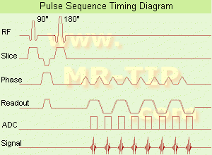
Echo Planar Imaging Timing Diagram (EPI) Echo planar imaging is one of the early magnetic resonance imaging sequences (also known as Intascan), used in applications like diffusion, perfusion, and functional magnetic resonance imaging. Other sequences acquire one k-space line at each phase encoding step. When the echo planar imaging acquisition strategy is used, the complete image is formed from a single data sample (all k-space lines are measured in one repetition time) of a gradient echo or spin echo sequence (see single shot technique) with an acquisition time of about 20 to 100 ms. The pulse sequence timing diagram illustrates an echo planar imaging sequence from spin echo type with eight echo train pulses. (See also Pulse Sequence Timing Diagram, for a description of the components.)
In case of a gradient echo based EPI sequence the initial part is very similar to a standard gradient echo sequence. By periodically fast reversing the readout or frequency encoding gradient, a train of echoes is generated.
EPI requires higher performance from the MRI scanner like much larger gradient amplitudes. The scan time is dependent on the spatial resolution required, the strength of the applied gradient fields and the time the machine needs to ramp the gradients.
In EPI, there is water fat shift in the phase encoding direction due to phase accumulations. To minimize water fat shift (WFS) in the phase direction fat suppression and a wide bandwidth (BW) are selected. On a typical EPI sequence, there is virtually no time at all for the flat top of the gradient waveform. The problem is solved by "ramp sampling" through most of the rise and fall time to improve image resolution.
The benefits of the fast imaging time are not without cost. EPI is relatively demanding on the scanner hardware, in particular on gradient strengths, gradient switching times, and receiver bandwidth. In addition, EPI is extremely sensitive to image artifacts and distortions.
spacer

• View the DATABASE results for 'Echo Planar Imaging' (19).Open this link in a new window


• View the NEWS results for 'Echo Planar Imaging' (1).Open this link in a new window.
 
Further Reading:
  Basics:
New Imaging Method Makes Brain Scans 7 Times Faster
Sunday, 9 January 2011   by www.dailytech.com    
Searchterm 'pulse sequence' was also found in the following service: 
spacer
Ultrasound  (1) Open this link in a new window
Gradient Motion Rephasing
 
(GMR) The application of strategic gradient pulses can compensate the objectionable spin phase effects of flow motion. That means the reducing of flow effects, e.g. gradient moment nulling of the first order of flow. The simplest velocity-compensated pulse sequence is the symmetrical second echo of a spin echo pulse sequence.
Gradient field changes can be configured in such a way that during an echo the magnetization signal vectors for all pixels have zero phase angle independent of velocities, accelerations etc. of the measured tissue. E.g. the adjustment to zero at the time TE of the net moments of the amplitude of the waveform of the magnetic field gradients with time. The zeroth moment is the area under the curve, the first moment is the 'center of gravity' etc. The aim is to minimize the phase shifts acquired by the transverse magnetization of excited nuclei moving along the gradients (including the effect of refocusing RF pulses), particularly for the reduction of image artifacts due to motion.
Also called Flow Compensation (FC), Motion Artifact Suppression Technique (MAST), Flow motion compression (STILL), Gradient Rephasing (GR), Shimadzu Motion Artifact Reduction Technique (SMART).
spacer

• View the DATABASE results for 'Gradient Motion Rephasing' (2).Open this link in a new window

 
Further Reading:
  Basics:
Motion Compensation in MR Imaging
   by ccn.ucla.edu    
MRI Resources 
MR Myelography - Lung Imaging - Mobile MRI Rental - Musculoskeletal and Joint MRI - Sequences - Claustrophobia
 
previous      21 - 25 (of 171)     next
Result Pages : [1]  [2 3 4 5 6 7 8 9 10 11 12 13 14 15 16 17 18 19 20 ... ]
 Random Page
 
Share This Page
FacebookTwitterLinkedIn

MR-TIP    
Community   
User
Pass
Forgot your UserID/Password ?    


MRI is trending to low field magnets :
reduced costs will lead to this change 
AI will close the gap to high field 
only in remote areas 
is only temporary 
never 

Look
      Ups


MR-TIP.com uses cookies! By browsing MR-TIP.com, you agree to our use of cookies.

Magnetic Resonance - Technology Information Portal
Member of SoftWays' Medical Imaging Group - MR-TIP • Radiology-TIP • Medical-Ultrasound-Imaging • 
Copyright © 2003 - 2024 SoftWays. All rights reserved. [ 21 November 2024]
Terms of Use | Privacy Policy | Advertising
 [last update: 2024-02-26 03:41:00]