Magnetic Resonance - Technology Information Portal Welcome to MRI Technology
Info
  Sheets

Out-
      side


 'sequence' 
SEARCH FOR    
 
  2 3 5 A B C D E F G H I J K L M N O P Q R S T U V W X Y Z
Result : Searchterm 'sequence' found in 30 terms [] and 347 definitions []
previous     91 - 95 (of 377)     next
Result Pages : [1 2 3 4 5 6]  [7 8 9 10 11 12 13 14 15 16 17 18 19 20 ... ]
Searchterm 'sequence' was also found in the following services: 
spacer
News  (28)  Resources  (10)  Forum  (106)  
 
Black Blood MRAForum -
related threadsInfoSheet: - Sequences - 
Intro, 
Overview, 
Types of, 
etc.MRI Resource Directory:
 - Cardiovascular Imaging -
 
With this magnetic resonance angiography technique flowing blood appears dark.
MR black blood techniques have been developed for cardiovascular imaging to improve segmentation of myocardium from the blood pool. Black blood MRA techniques decrease the signal from blood with reference to the myocardium and make it easier to perform cardiac chamber segmentation.
ECG gated spin echo sequences with presaturation pulses for magnetization preparation will show strong intravascular signal loss due to flow effects when appropriate imaging conditions including spatial presaturation are used. The sequence use the flow void effect as blood passes rapidly through the selected slice.
For dark blood preparation, a pair of nonselective and selective 180° inversion pulses are used, followed by a long inversion time to null signal from inflowing blood. A second selective inversion pulse can also be applied with short inversion time to null the fat signal. These in cardiac imaging used black blood techniques are referred to as double inversion recovery T1 measurement turbo spin echo or fast spin echo, and double-inversion recovery STIR.
 
Images, Movies, Sliders:
 Normal Dual Inversion Fast Spin-echo  Open this link in a new window
      

Courtesy of  Robert R. Edelman

 
spacer
 
• Related Searches:
    • Double Inversion Recovery T1 Measurement
    • Inversion
    • Blood Flow Imaging
    • Displacement Encoding with Stimulated Echoes
    • Magnetic Resonance Angiography MRA
MRI Resources 
Societies - Functional MRI - Contrast Enhanced MRI - Services and Supplies - Brain MRI - MR Guided Interventions
 
Black Boundary ArtifactInfoSheet: - Artifacts - 
Case Studies, 
Reduction Index, 
etc.MRI Resource Directory:
 - Artifacts -
 
Quick Overview
Please note that there are different common names for this artifact.
Artifact Information
NAME
Black boundary, dark boundary, contour, chemical shift, relief
DESCRIPTION
Black contours at boundaries
Black boundary artifacts are black lines following voxels where both water and fat protons are present in the same voxel. This artifact arise along the boundary of organs or tissues perpendicular to the frequency encoding direction, and occurs preferentially in gradient echo sequences with out of phase echo times.
mri safety guidance
Image Guidance
Fat suppression techniques eliminate this artifact. For artifact reducing helps a smaller water fat shift (high bandwidth), a higher matrix or/and an in phase TE.

See also Chemical Shift Artifact.
spacer

• View the DATABASE results for 'Black Boundary Artifact' (4).Open this link in a new window

 
Further Reading:
  Basics:
What is chemical shift artefact? Why does it occur? How many Hz at 1.5 T?
   by www.revisemri.com    
MRI Resources 
MRI Technician and Technologist Schools - RIS - MRI Centers - Shielding - NMR - Equipment
 
Breath Hold ImagingMRI Resource Directory:
 - Abdominal Imaging -
 
Breath hold imaging in MRI is a technique with one ore more stoppage of breathing during the sequence and require therefore a short scan time. Breath hold techniques are used with fast gradient echo sequences in thoracic or abdominal regions with much respiratory movement.
Breath hold cine MRI techniques are used in cardiovascular imaging and provide detailed views of the beating heart in different cardiac axes.
Breath hold imaging requires the full cooperation of the patient, caused by usual MRI scan times from 15 to 20 sec.. In some cases breath holding can be practiced outside the MRI scanner to improve patient cooperation with the examination. Shorter scan times e.g. by parallel imaging techniques, or the administration of oxygen can help the patient to hold the breath during the scan.
See also Abdominal Imaging.
 
Images, Movies, Sliders:
 MRI Upper Abdomen T1 with Contrast  Open this link in a new window
 Normal Dual Inversion Fast Spin-echo  Open this link in a new window
 Anatomic Imaging of the Lungs  Open this link in a new window
 
spacer

• View the DATABASE results for 'Breath Hold Imaging' (7).Open this link in a new window

 
Further Reading:
  News & More:
The Effects of Breathing Motion on DCE-MRI Images: Phantom Studies Simulating Respiratory Motion to Compare CAIPIRINHA-VIBE, Radial-VIBE, and Conventional VIBE
Tuesday, 7 February 2017   by www.kjronline.org    
Controlling patient's breathing makes cardiac MRI more accurate
Friday, 13 May 2016   by www.upi.com    
Accurate T1 Quantification Using a Breath-hold Inversion Recovery TrueFISP Sequence
2003   by rsna2003.rsna.org    
Searchterm 'sequence' was also found in the following services: 
spacer
News  (28)  Resources  (10)  Forum  (106)  
 
CHORUS 1.5Tâ„¢InfoSheet: - Devices -
Intro, 
Types of Magnets, 
Overview, 
etc.MRI Resource Directory:
 - Devices -
 
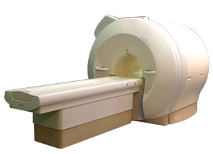
www.isoltech.co.kr/english/product/default.htm 'Next generation MRI system 1.5T CHORUS developed by ISOL Technology is optimized for both clinical diagnostic imaging and for research development.
CHORUS offers the complete range of feature oriented advanced imaging techniques- for both clinical routine and research. The compact short bore magnet, the patient friendly design and the gradient technology make the innovation to new degree of perfection in magnetic resonance.'
Device Information and Specification
CLINICAL APPLICATION
Whole body
CONFIGURATION
Short bore
Head, C-spine, L-spine, TMJ, Knee, Shoulder, General purpose, Phased Array System: 4 digital receiver channels (Up to 12 channels)
Optional
SYNCHRONIZATION
ECG/peripheral: Optional/yes, respiratory gating
PULSE SEQUENCES
Spin Echo, Gradient Echo, Fast Spin Echo, Inversion Recovery (STIR, Fluid Attenuated Inversion Recovery), FLASH, FISP, PSIF, Turbo Flash ( MPRAGE ),TOF MR Angiography, Standard echo planar imaging package (SE-EPI, GE-EPI), Optional: Advanced P.A. Imaging Package (up to 4 ch.), Advanced echo planar imaging package, Single Shot and Diffusion Weighted EPI, IR/FLAIR EPI
IMAGING MODES
2D/3D, Travelling Sat, Multi-Slab 3D, MTC and TONE Pulse Sequence, Fat/Water Suppression, Presaturation (up to 6 bands), Flow Compensation using GMR pulse, Multi-Slice, Multi-Group Imaging
SINGLE/MULTI SLICE
Image reconstruction time (2562 ) : 0.02 s
FOV
40 cm
BORE DIAMETER
or W x H
58 cm diameter (patient)
MAGNET WEIGHT
4050 kg
H*W*D
233 x 206 x 160 cm
COOLING SYSTEM TYPE
Water-cooled coil and air-cooled amplifier
CRYOGEN USE
0.07 L/hr helium
STRENGTH
20 mT/m (Upto 27 mT/m)
5-GAUSS FRINGE FIELD
2.5 m / 3.8 m
Passive and active
spacer

• View the DATABASE results for 'CHORUS 1.5T™' (2).Open this link in a new window

MRI Resources 
RIS - Breast Implant - Online Books - Shielding - Stimulator pool - MRI Reimbursement
 
Cardiac Gating
 
This method synchronize the heartbeat with the beginning of the TR, whereat the r wave is used as the trigger. Cardiac gating times the acquisition of MR data to physiological motion in order to minimize motion artifacts. ECG gating techniques are useful whenever data acquisition is too slow to occur during a short fraction of the cardiac cycle.
Image blurring due to cardiac-induced motion occurs for imaging times of above approximately 50 ms in systole, while for imaging during diastole the critical time is of the order of 200-300 ms. The acquisition of an entire image in this time is only possible with using ultrafast MR imaging techniques. If a series of images using cardiac gating or real-time echo planar imaging EPI are acquired over the entire cardiac cycle, pixel-wise velocity and vascular flow can be obtained.
In simple cardiac gating, a single image line is acquired in each cardiac cycle. Lines for multiple images can then be acquired successively in consecutive gate intervals. By using the standard multiple slice imaging and a spin echo pulse sequence, a number of slices at different anatomical levels is obtained. The repetition time (TR) during a ECG-gated acquisition equals the RR interval, and the RR interval defines the minimum possible repetition time (TR). If longer TRs are required, multiple integers of the RR interval can be selected. When using a gradient echo pulse sequence, multiple phases of a single anatomical level or multiple slices at different anatomical levels can be acquired over the cardiac cycle.
Also called cardiac triggering.
 
Images, Movies, Sliders:
 Cardiac Infarct Short Axis Cine Overview  Open this link in a new window
    

Courtesy of  Robert R. Edelman
 Infarct 4 Chamber Cine  Open this link in a new window
    

Courtesy of  Robert R. Edelman
 
spacer

• View the DATABASE results for 'Cardiac Gating' (15).Open this link in a new window

 
Further Reading:
  Basics:
Cardiac MRI - Technical Aspects Primer
Wednesday, 7 August 2002
Electrocardiogram in an MRI Environment: Clinical Needs, Practical Considerations, Safety Implications, Technical Solutions and Future Directions
Wednesday, 25 January 2012   by cdn.intechopen.com    
Motion-compensation of Cardiac Perfusion MRI using a Statistical Texture Ensemble(.pdf)
June 2003   by www.imm.dtu.dk    
MRI Resources 
Bioinformatics - IR - Open Directory Project - Anatomy - Equipment - Cardiovascular Imaging
 
previous      91 - 95 (of 377)     next
Result Pages : [1 2 3 4 5 6]  [7 8 9 10 11 12 13 14 15 16 17 18 19 20 ... ]
 Random Page
 
Share This Page
FacebookTwitterLinkedIn

MR-TIP    
Community   
User
Pass
Forgot your UserID/Password ?    


New acceleration techniques will :
reduce scan times 
cause artifacts 
increase expenses 
be useful if you have a lot of experience 
doesn't do much 
never heard of 

Look
      Ups


MR-TIP.com uses cookies! By browsing MR-TIP.com, you agree to our use of cookies.

Magnetic Resonance - Technology Information Portal
Member of SoftWays' Medical Imaging Group - MR-TIP • Radiology-TIP • Medical-Ultrasound-Imaging • 
Copyright © 2003 - 2024 SoftWays. All rights reserved. [ 4 December 2024]
Terms of Use | Privacy Policy | Advertising
 [last update: 2024-02-26 03:41:00]