Magnetic Resonance - Technology Information Portal Welcome to MRI Technology
Info
  Sheets

Out-
      side


 't2' 
SEARCH FOR    
 
  2 3 5 A B C D E F G H I J K L M N O P Q R S T U V W X Y Z
Result : Searchterm 't2' found in 8 terms [] and 150 definitions []
previous     11 - 15 (of 158)     next
Result Pages : [1 2]  [3 4 5 6 7 8 9 10 11 12 13 14 15 16 17 18 19 20 ... ]
Searchterm 't2' was also found in the following services: 
spacer
News  (28)  Resources  (5)  Forum  (67)  
 
Gradient Echo SequenceForum -
related threadsInfoSheet: - Sequences - 
Intro, 
Overview, 
Types of, 
etc.
 
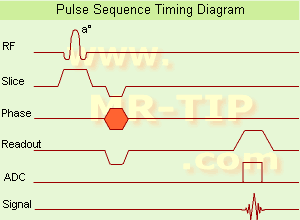
Gradient Echo Sequence Timing Diagram (GRE - sequence) A gradient echo is generated by using a pair of bipolar gradient pulses. In the pulse sequence timing diagram, the basic gradient echo sequence is illustrated. There is no refocusing 180° pulse and the data are sampled during a gradient echo, which is achieved by dephasing the spins with a negatively pulsed gradient before they are rephased by an opposite gradient with opposite polarity to generate the echo.
See also the Pulse Sequence Timing Diagram. There you will find a description of the components.
The excitation pulse is termed the alpha pulse α. It tilts the magnetization by a flip angle α, which is typically between 0° and 90°. With a small flip angle there is a reduction in the value of transverse magnetization that will affect subsequent RF pulses. The flip angle can also be slowly increased during data acquisition (variable flip angle: tilt optimized nonsaturation excitation). The data are not acquired in a steady state, where z-magnetization recovery and destruction by ad-pulses are balanced. However, the z-magnetization is used up by tilting a little more of the remaining z-magnetization into the xy-plane for each acquired imaging line.
Gradient echo imaging is typically accomplished by examining the FID, whereas the read gradient is turned on for localization of the signal in the readout direction. T2* is the characteristic decay time constant associated with the FID. The contrast and signal generated by a gradient echo depend on the size of the longitudinal magnetization and the flip angle. When α = 90° the sequence is identical to the so-called partial saturation or saturation recovery pulse sequence. In standard GRE imaging, this basic pulse sequence is repeated as many times as image lines have to be acquired. Additional gradients or radio frequency pulses are introduced with the aim to spoil to refocus the xy-magnetization at the moment when the spin system is subject to the next α pulse.
As a result of the short repetition time, the z-magnetization cannot fully recover and after a few initial α pulses there is an equilibrium established between z-magnetization recovery and z-magnetization reduction due to the α pulses.
Gradient echoes have a lower SAR, are more sensitive to field inhomogeneities and have a reduced crosstalk, so that a small or no slice gap can be used. In or out of phase imaging depending on the selected TE (and field strength of the magnet) is possible. As the flip angle is decreased, T1 weighting can be maintained by reducing the TR. T2* weighting can be minimized by keeping the TE as short as possible, but pure T2 weighting is not possible. By using a reduced flip angle, some of the magnetization value remains longitudinal (less time needed to achieve full recovery) and for a certain T1 and TR, there exist one flip angle that will give the most signal, known as the "Ernst angle".
Contrast values:
PD weighted: Small flip angle (no T1), long TR (no T1) and short TE (no T2*)
T1 weighted: Large flip angle (70°), short TR (less than 50ms) and short TE
T2* weighted: Small flip angle, some longer TR (100 ms) and long TE (20 ms)

Classification of GRE sequences can be made into four categories:
See also Gradient Recalled Echo Sequence, Spoiled Gradient Echo Sequence, Refocused Gradient Echo Sequence, Ultrafast Gradient Echo Sequence.
 
Images, Movies, Sliders:
 MRI Liver In Phase  Open this link in a new window
    
 MRI Liver Out Of Phase  Open this link in a new window
    
 MVP Parasternal  Open this link in a new window
 Breast MRI Images T1 Pre - Post Contrast  Open this link in a new window
 Circle of Willis, Time of Flight, MIP  Open this link in a new window
    
SlidersSliders Overview

 
spacer
 
• Related Searches:
    • MRI History
    • Pulse Sequence Timing Diagram
    • Abdominal Imaging
    • Contrast
    • Ultrafast Gradient Echo Sequence
 
Further Reading:
  Basics:
Enhanced Fast GRadient Echo 3-Dimensional (efgre3D) or THRIVE
   by www.mri.tju.edu    
  News & More:
MRI evaluation of fatty liver in day to day practice: Quantitative and qualitative methods
Wednesday, 3 September 2014   by www.sciencedirect.com    
T1rho-prepared balanced gradient echo for rapid 3D T1rho MRI
Monday, 1 September 2008   by www.ncbi.nlm.nih.gov    
MRI Resources 
Used and Refurbished MRI Equipment - MRI Technician and Technologist Career - Musculoskeletal and Joint MRI - Calculation - MRI Technician and Technologist Jobs - Anatomy
 
Contrast Enhanced Gradient Echo SequenceInfoSheet: - Sequences - 
Intro, 
Overview, 
Types of, 
etc.
 
Contrast enhanced GRE sequences provide T2 contrast but have a relatively poor SNR. Repetitive RF pulses with small flip angles together with appropriate gradient profiles lead to the superposition of two resonance signals.
The first signal is due to the free induction decay FID observed after the first and all ensuing RF excitations.
The second is a resonance signal obtained as a result of a spin echo generated by the second and all addicted RF-pulses.
Hence it is absent after the first excitation, it is a result of the free induction decay of the second to last RF-excitation and has a TE, which is almost 2TR. For this echo to occur the gradients have to be completely symmetrical relative to the half time between two RF-pulses, a condition that makes it difficult to integrate this pulse sequence into a multiple slice imaging technique. The second signal not only contains echo contributions from free induction decay, but obviously weakened by T2-decay. Since the echo is generated by a RF-pulse, it is truly T2 rather than T2* weighted. Correspondingly it is also less sensitive to susceptibility changes and field inhomogeneities.
Companies use different acronyms to describe certain techniques.
Different terms (see also acronyms) for these gradient echo pulse sequences:
CE-FAST Contrast Enhanced Fourier Acquired Steady State,
CE-FFE Contrast Enhanced Fast Field Echo,
CE-GRE Contrast Enhanced Gradient-Echo,
DE-FGR Driven Equilibrium FGR,
FADE FASE Acquisition Double Echo,
PSIF Reverse Fast Imaging with Steady State Precession,
SSFP Steady State Free Precession,
T2 FFE Contrast Enhanced Fast Field Echo (T2 weighted).

In this context, 'contrast enhanced' refers to the pulse sequence, it does not mean enhancement with a contrast agent.
spacer

• View the DATABASE results for 'Contrast Enhanced Gradient Echo Sequence' (4).Open this link in a new window

MRI Resources 
Pathology - Equipment - Chemistry - Coils - Journals - Universities
 
Perfusion ImagingForum -
related threadsInfoSheet: - Sequences - 
Intro, 
Overview, 
Types of, 
etc.
 
(PWI - Perfusion Weighted Imaging) Perfusion MRI techniques (e.g. PRESTO - Principles of Echo Shifting using a Train of Observations) are sensitive to microscopic levels of blood flow. Contrast enhanced relative cerebral blood volume (rCBV) is the most used perfusion imaging. Both, the ready availability and the T2* susceptibility effects of gadolinium, rather than the T1 shortening effects make gadolinium a suitable agent for use in perfusion imaging. Susceptibility here refers to the loss of MR signal, most marked on T2* (gradient echo)-weighted and T2 (spin echo)-weighted sequences, caused by the magnetic field-distorting effects of paramagnetic substances.
T2* perfusion uses dynamic sequences based on multi or single shot techniques. The T2* (T2) MRI signal drop within or across a brain region is caused by spin dephasing during the rapid passage of contrast agent through the capillary bed. The signal decrease is used to compute the relative perfusion to that region. The bolus through the tissue is only a few seconds, high temporal resolution imaging is required to obtain sequential images during the wash in and wash out of the contrast material and therefore, resolve the first pass of the tracer. Due to the high temporal resolution, processing and calculation of hemodynamic maps are available (including mean transit time (MTT), time to peak (TTP), time of arrival (T0), negative integral (N1) and index.
An important neuroradiological indication for MRI is the evaluation of incipient or acute stroke via perfusion and diffusion imaging. Diffusion imaging can demonstrate the central effect of a stroke on the brain, whereas perfusion imaging visualizes the larger 'second ring' delineating blood flow and blood volume. Qualitative and in some instances quantitative (e.g. quantitative imaging of perfusion using a single subtraction) maps of regional organ perfusion can thus be obtained.
Echo planar and potentially echo volume techniques together with appropriate computing power offer real time images of dynamic variations in water characteristics reflecting perfusion, diffusion, oxygenation (see also Oxygen Mapping) and flow.
Another type of perfusion MR imaging allows the evaluation of myocardial ischemia during pharmacologic stress. After e.g., adenosine infusion, multiple short axis views (see cardiac axes) of the heart are obtained during the administration of gadolinium contrast. Ischemic areas show up as areas of delayed and diminished enhancement. The MRI stress perfusion has been shown to be more accurate than nuclear SPECT exams. Myocardial late enhancement and stress perfusion imaging can also be performed during the same cardiac MRI examination.
 
Images, Movies, Sliders:
 Normal Lung Gd Perfusion MRI  Open this link in a new window
      

Courtesy of  Robert R. Edelman

 Left Circumflex Ischemia First-pass Contrast Enhancement  Open this link in a new window
 
Radiology-tip.comradPerfusion Scintigraphy
spacer
Medical-Ultrasound-Imaging.comBolus Injection
spacer

• View the DATABASE results for 'Perfusion Imaging' (16).Open this link in a new window


• View the NEWS results for 'Perfusion Imaging' (3).Open this link in a new window.
 
Further Reading:
  Basics:
CHAPTER 55: Ischemia
2003
EVALUATION OF HUMAN STROKE BY MR IMAGING
2000
  News & More:
Non-invasive diagnostic procedures for suspected CHD: Search reveals informative evidence
Wednesday, 8 July 2020   by medicalxpress.co    
Implementation of Dual-Source RF Excitation in 3 T MR-Scanners Allows for Nearly Identical ADC Values Compared to 1.5 T MR Scanners in the Abdomen
Wednesday, 29 February 2012   by www.plosone.org    
Motion-compensation of Cardiac Perfusion MRI using a Statistical Texture Ensemble(.pdf)
June 2003   by www.imm.dtu.dk    
Turbo-FLASH Based Arterial Spin Labeled Perfusion MRI at 7 T
Thursday, 20 June 2013   by www.plosone.org    
Measuring Cerebral Blood Flow Using Magnetic Resonance Imaging Techniques
1999   by www.stanford.edu    
Vascular Filters of Functional MRI: Spatial Localization Using BOLD and CBV Contrast
Searchterm 't2' was also found in the following services: 
spacer
News  (28)  Resources  (5)  Forum  (67)  
 
T1 WeightedInfoSheet: - Sequences - 
Intro, 
Overview, 
Types of, 
etc.
 
Every tissue in the human body has its own T1 and T2 value. This term is used to indicate an image where most of the contrast between tissues is due to differences in the T1 value.
This term may be misleading in that the potentially important effects of tissue density differences and the range of tissue T1 values are ignored.
If the machine parameters are chosen, so that TR less than T1 (typically under 500 ms) and TE less than T2 (typically under 30 ms), a power series expansion of the exponential functions and then neglecting second and higher order terms yields
Mxy = Mxy0 TR/T1
thus the expression becomes independent of T2 and yields the condition for T1 weighting. Therefore a T1 contrast is approached by imaging with a short TR, compared to the longest tissue T1 of interest and short TE, compared to tissue T2 (to reduce T2 contributions to image contrast). Due to the wide range of T1 and T2 and tissue density values that can be found in the body, an image that is T1 weighted for some tissues may not be so for others.
Lesions with short T1 are (bright in T1 weighted sequences):
fat (lipoma, dermoid)
sub-acute haemorrhage (metHb)
paramagnetic agent (Gd, pituitary)
protein-containing fluid (colloid cyst)
metastatic melanoma (melanotic).
 
Images, Movies, Sliders:
 MRI Orbita T1  Open this link in a new window
    
 MRI Liver In Phase  Open this link in a new window
    
 Sagittal Knee MRI Images T1 Weighted  Open this link in a new window
 MRI - Anatomic Imaging of the Ankle 2  Open this link in a new window
    
SlidersSliders Overview

 
spacer

• View the DATABASE results for 'T1 Weighted' (56).Open this link in a new window

 
Further Reading:
  Basics:
A paired dataset of T1- and T2-weighted MRI at 3 Tesla and 7 Tesla
Thursday, 27 July 2023   by www.nature.com    
A practical guideline for T1 reconstruction from various flip angles in MRI
Saturday, 1 October 2016   by journals.sagepub.com    
Accurate T1 Quantification Using a Breath-hold Inversion Recovery TrueFISP Sequence
2003   by rsna2003.rsna.org    
  News & More:
T1rho-prepared balanced gradient echo for rapid 3D T1rho MRI
Monday, 1 September 2008   by www.ncbi.nlm.nih.gov    
Possible New MRI Marker for Multiple Sclerosis Progression
Wednesday, 29 August 2007   by www.medpagetoday.com    
T1-weighted Phase Sensitive Inversion Recovery for Imaging Multiple Sclerosis Lesions in the Cervical Spinal Cord(.pdf)
   by www.healthcare.siemens.com    
MRI Resources 
Knee MRI - MRA - Safety pool - Pacemaker - Raman Spectroscopy - Spectroscopy
 
Coherent Gradient EchoInfoSheet: - Sequences - 
Intro, 
Overview, 
Types of, 
etc.MRI Resource Directory:
 - Sequences -
 
Coherent gradient echo sequences can measure the free induction decay (FID), generated just after each excitation pulse or the echo formed prior to the next pulse. Coherent gradient echo sequences are very sensitive to magnetic field inhomogeneity. An alternative to spoiling is to incorporate residual transverse magnetization directly into the longitudinal steady state. These GRE sequences use a refocusing gradient in the phase encoding direction during the end module to maximize remaining transverse (xy) magnetization at the time when the next excitation is due, while the other two gradients are, in any case, balanced.
When the next excitation pulse is sent into the system with an opposed phase, it tilts the magnetization in the -a direction. As a result the z-magnetization is again partly tilted into the xy-plane, while the remaining xy-magnetization is tilted partly into the z-direction.
A fully refocused sequence with a properly selected and uniform f would yield higher signal, especially for tissues with long T2 relaxation times (high water content) so it is used in angiographic, myelographic or arthrographic examinations and is used for T2* weighting. The repetition time for this sequence has to be short. With short TR, coherent GE is also useable for breath hold and 3D technique. If the repetition time is about 200 msec there's no difference between spoiled or unspoiled GE. T1 weighting is better with spoiled techniques.
The common types include GRASS, FISP, FAST, and FFE.
The T2* component decreases with long TR and short TE. The T1 time is controlled by flip angle. The common TR is less than 50 ms and the common TE less than 15 ms
Other types have stronger T2 dependence but lower SNR. They include SSFP, CE-FAST, PSIF, and CE-FFE-T2.
Examples of fully refocused FID sequences are TrueFISP, bFFE and bTFE.
spacer

• View the DATABASE results for 'Coherent Gradient Echo' (6).Open this link in a new window

MRI Resources 
Journals - Health - Safety Training - Abdominal Imaging - Stimulator pool - General
 
previous      11 - 15 (of 158)     next
Result Pages : [1 2]  [3 4 5 6 7 8 9 10 11 12 13 14 15 16 17 18 19 20 ... ]
 Random Page
 
Share This Page
FacebookTwitterLinkedIn

MR-TIP    
Community   
User
Pass
Forgot your UserID/Password ?    


Next big thing in MRI will be :
AI 
remote operator 
personalized protocols 
helium-free 
molecular MRI 
portable MRI 

Look
      Ups


MR-TIP.com uses cookies! By browsing MR-TIP.com, you agree to our use of cookies.

Magnetic Resonance - Technology Information Portal
Member of SoftWays' Medical Imaging Group - MR-TIP • Radiology-TIP • Medical-Ultrasound-Imaging • 
Copyright © 2003 - 2024 SoftWays. All rights reserved. [ 21 November 2024]
Terms of Use | Privacy Policy | Advertising
 [last update: 2024-02-26 03:41:00]