Magnetic Resonance - Technology Information Portal Welcome to MRI Technology
Info
  Sheets

Out-
      side


 'Phase' 
SEARCH FOR    
 
  2 3 5 A B C D E F G H I J K L M N O P Q R S T U V W X Y Z
Result : Searchterm 'Phase' found in 35 terms [] and 251 definitions []
previous     51 - 55 (of 286)     next
Result Pages : [1 2 3 4 5 6 7]  [8 9 10 11 12 13 14 15 16 17 18 19 20 ... ]
Searchterm 'Phase' was also found in the following services: 
spacer
News  (25)  Resources  (11)  Forum  (27)  
 
iMotion™ 1.5 Tesla MagnetInfoSheet: - Devices -
Intro, 
Types of Magnets, 
Overview, 
etc.MRI Resource Directory:
 - Devices -
 
www.imris.com/ The mobile intraoperative iMotion system produces real-time images used for MR guided surgery and offers functional magnetic resonance imaging, MR spectroscopy, perfusion imaging, and diffusion weighted imaging capabilities.
The iMotion 1.5 T magnet moves to the patient, gliding in and out of place as needed, without affecting surgical, anesthetic, and nursing management.

See also Intraoperative Magnetic Resonance Imaging, MR Guided Interventions.
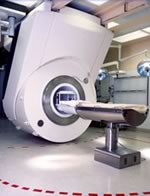
Device Information and Specification
CLINICAL APPLICATION
Whole body
CONFIGURATION
Mobile compact
Whole body, intra-operative head, neck volume, atlas head//neck vascular quadrature phased array, spine quadrature, C/T/L spine phased array, small joint, large joint, TMJ bilateral, shoulder phased array, extremity quadrature volume, wrist, hand quadrature, general purpose flexible, pelvis/abdomen phased array, body quadrature, phased array flexible, breast bilateral
SYNCHRONIZATION
Standard cardiac gating, ECG/peripheral, respiratory gating
PULSE SEQUENCES
SE, IR, TSE, EPI, Diffusion, Perfusion, Angiography
IMAGING MODES
Localizer, single slice, multislice, volume
Active
spacer
Searchterm 'Phase' was also found in the following services: 
spacer
Radiology  (17) Open this link in a new windowUltrasound  (77) Open this link in a new window
Bipolar Gradient Pulse
 
Bipolar gradients are two gradients with the same magnitude but opposite gradient direction. A bipolar gradient pulse is produced if one of the bipolar gradients is switched e.g., in negative direction and then switched in the opposite direction for an equivalent amount of time.
Bipolar gradients are used e.g. in phase contrast and diffusion weighted sequences. A bipolar gradient pulse pair produces a phase shift, which depends on the velocity component along this gradient. Motion along a bipolar gradient pulse pair results in a flow-induced phase shift of the transverse magnetization. The bipolar gradient pulse pair will not affect stationary spins. The amount of phase shifts depends on the area of each gradient pulse, and distance between the pulses. An echo occurring after such a gradient is flow compensated for velocity. A slight shift in the balance of this gradient will introduce a defined flow sensitivity of the pulse sequence.
 
Images, Movies, Sliders:
 PCA-MRA 3D Brain Venography Colored MIP  Open this link in a new window


spacer

• View the DATABASE results for 'Bipolar Gradient Pulse' (7).Open this link in a new window

MRI Resources 
Software - Collections - Liver Imaging - Health - Absorption and Emission - Online Books
 
Black Boundary ArtifactInfoSheet: - Artifacts - 
Case Studies, 
Reduction Index, 
etc.MRI Resource Directory:
 - Artifacts -
 
Quick Overview
Please note that there are different common names for this artifact.
Artifact Information
NAME
Black boundary, dark boundary, contour, chemical shift, relief
DESCRIPTION
Black contours at boundaries
Black boundary artifacts are black lines following voxels where both water and fat protons are present in the same voxel. This artifact arise along the boundary of organs or tissues perpendicular to the frequency encoding direction, and occurs preferentially in gradient echo sequences with out of phase echo times.
mri safety guidance
Image Guidance
Fat suppression techniques eliminate this artifact. For artifact reducing helps a smaller water fat shift (high bandwidth), a higher matrix or/and an in phase TE.

See also Chemical Shift Artifact.
spacer

• View the DATABASE results for 'Black Boundary Artifact' (4).Open this link in a new window

 
Further Reading:
  Basics:
What is chemical shift artefact? Why does it occur? How many Hz at 1.5 T?
   by www.revisemri.com    
Searchterm 'Phase' was also found in the following services: 
spacer
News  (25)  Resources  (11)  Forum  (27)  
 
Dual Echo Fast Gradient EchoInfoSheet: - Sequences - 
Intro, 
Overview, 
Types of, 
etc.
 
(DE FGRE, Dual/FFE, DE FFE) Simultaneously acquired in and out of phase TE gradient echo images. To quantitatively measure the signal intensity differences between out of phase and in phase images the parameters should be the same except for the TE.
The chemical shift artifact appearing on the out-of-phase image allows for the detection of lipids in the liver or adrenal gland, such as diffuse fatty infiltration, focal fatty infiltration, focal fatty sparing, benign adrenocortical masses and intracellular lipids within a hepatocellar neoplasm, where spin echo and fat suppression techniques are not as sensitive. Specific pathologies that have been reported include liver lipoma, angiomyolipoma, myelolipoma, metastatic liposarcoma, teratocarcinoma, melanoma, haemorrhagic neoplasm and metastatic choriocarcinoma.
 
Images, Movies, Sliders:
 MRI Liver In Phase  Open this link in a new window
    
 MRI Liver Out Of Phase  Open this link in a new window
    
 
spacer

• View the DATABASE results for 'Dual Echo Fast Gradient Echo' (2).Open this link in a new window

 
Further Reading:
  News & More:
Adrenal Myelolipoma
Tuesday, 19 June 2001   by www.emedicine.com    
Searchterm 'Phase' was also found in the following services: 
spacer
Radiology  (17) Open this link in a new windowUltrasound  (77) Open this link in a new window
Echo Offset
 
Echo offset is the time setting of spin echo and gradient echo to be not coincident and to generate phase differences between different spectral line signals (e.g., water and fat). The echo offset is the product of the frequency line difference and the time difference (TD) in the echo times and is equal to the magnitude of the result of the phase difference between two spectral lines. Phases may not change linearly with echo offset time in the presence of a large field inhomogeneity. An echo offset excitation pulse sequence can be used in the magnetic field mapping method, to generate maps from which the standard deviation of the phase difference can be calculated.
spacer
 
Further Reading:
  News & More:
T1rho-prepared balanced gradient echo for rapid 3D T1rho MRI
Monday, 1 September 2008   by www.ncbi.nlm.nih.gov    
A short-TR single-echo spin-echo breath-hold method for assessing liver T2
Sunday, 10 December 2023   by link.springer.com    
MRI Resources 
Devices - Case Studies - DICOM - Online Books - Developers - Spectroscopy pool
 
previous      51 - 55 (of 286)     next
Result Pages : [1 2 3 4 5 6 7]  [8 9 10 11 12 13 14 15 16 17 18 19 20 ... ]
 Random Page
 
Share This Page
FacebookTwitterLinkedIn

MR-TIP    
Community   
User
Pass
Forgot your UserID/Password ?    


MRI is trending to low field magnets :
reduced costs will lead to this change 
AI will close the gap to high field 
only in remote areas 
is only temporary 
never 

Look
      Ups


MR-TIP.com uses cookies! By browsing MR-TIP.com, you agree to our use of cookies.

Magnetic Resonance - Technology Information Portal
Member of SoftWays' Medical Imaging Group - MR-TIP • Radiology-TIP • Medical-Ultrasound-Imaging • 
Copyright © 2003 - 2024 SoftWays. All rights reserved. [ 4 May 2024]
Terms of Use | Privacy Policy | Advertising
 [last update: 2024-02-26 03:41:00]