Magnetic Resonance - Technology Information Portal Welcome to MRI Technology
Info
  Sheets

Out-
      side


 'Phase' 
SEARCH FOR    
 
  2 3 5 A B C D E F G H I J K L M N O P Q R S T U V W X Y Z
Result : Searchterm 'Phase' found in 35 terms [] and 251 definitions []
previous     56 - 60 (of 286)     next
Result Pages : [1 2 3 4 5 6 7]  [8 9 10 11 12 13 14 15 16 17 18 19 20 ... ]
Searchterm 'Phase' was also found in the following services: 
spacer
News  (25)  Resources  (11)  Forum  (27)  
 
Echo Planar ImagingInfoSheet: - Sequences - 
Intro, 
Overview, 
Types of, 
etc.MRI Resource Directory:
 - Sequences -
 
Echo Planar Imaging Timing Diagram (EPI) Echo planar imaging is one of the early magnetic resonance imaging sequences (also known as Intascan), used in applications like diffusion, perfusion, and functional magnetic resonance imaging. Other sequences acquire one k-space line at each phase encoding step. When the echo planar imaging acquisition strategy is used, the complete image is formed from a single data sample (all k-space lines are measured in one repetition time) of a gradient echo or spin echo sequence (see single shot technique) with an acquisition time of about 20 to 100 ms. The pulse sequence timing diagram illustrates an echo planar imaging sequence from spin echo type with eight echo train pulses. (See also Pulse Sequence Timing Diagram, for a description of the components.)
In case of a gradient echo based EPI sequence the initial part is very similar to a standard gradient echo sequence. By periodically fast reversing the readout or frequency encoding gradient, a train of echoes is generated.
EPI requires higher performance from the MRI scanner like much larger gradient amplitudes. The scan time is dependent on the spatial resolution required, the strength of the applied gradient fields and the time the machine needs to ramp the gradients.
In EPI, there is water fat shift in the phase encoding direction due to phase accumulations. To minimize water fat shift (WFS) in the phase direction fat suppression and a wide bandwidth (BW) are selected. On a typical EPI sequence, there is virtually no time at all for the flat top of the gradient waveform. The problem is solved by "ramp sampling" through most of the rise and fall time to improve image resolution.
The benefits of the fast imaging time are not without cost. EPI is relatively demanding on the scanner hardware, in particular on gradient strengths, gradient switching times, and receiver bandwidth. In addition, EPI is extremely sensitive to image artifacts and distortions.
spacer
 
• Related Searches:
    • Gradient Echo
    • Functional Magnetic Resonance Imaging
    • Diffusion Weighted Imaging
    • Sensitivity Encoding
    • K-Space Trajectory
 
Further Reading:
  Basics:
New Imaging Method Makes Brain Scans 7 Times Faster
Sunday, 9 January 2011   by www.dailytech.com    
Searchterm 'Phase' was also found in the following services: 
spacer
Radiology  (17) Open this link in a new windowUltrasound  (77) Open this link in a new window
Flow ArtifactInfoSheet: - Artifacts - 
Case Studies, 
Reduction Index, 
etc.MRI Resource Directory:
 - Artifacts -
 
Quick Overview
Please note that there are different common names for this artifact.
Artifact Information
DESCRIPTION
Vascular ghosts (ghosting artifact), anomalous intensities in images
REASON
Movement of body fluids
HELP
Flow compensation, presaturation, triggering
Flow effects in MRI produce a range of artifacts, e.g. intravascular signal void by time of flight effects; turbulent dephasing and first echo dephasing, caused by flowing blood.
Through movement of the hydrogen nuclei (e.g. blood flow), there is a location change between the time these nuclei experience a radio frequency pulse and the time the emitted signal is received (because the repetition time is asynchronous with the pulsatile flow).
The blood flow occasionally produces intravascular high signal intensities due to flow related enhancement, even echo rephasing and diastolic pseudogating. The pulsatile laminar flow within vessels often produces a complex multilayered band that usually propagates outside the head in the phase encoded direction. Blood flow artifacts should be considered as a special subgroup of motion artifacts.
mri safety guidance
Image Guidance
Artifacts can be reduced by reduction of phase shifts with flow compensation (gradient moment nulling), suppression of the blood signal with saturation pulses parallel to the slices, synchronization of the imaging sequence with the heart cycle (cardiac triggering) or can be flipped 90° by swapping the phase//frequency encoding directions.

See also Flow Related Enhancement and Flow Effects.
 
Images, Movies, Sliders:
 Knee MRI Sagittal T1 003  Open this link in a new window
 
spacer

• View the DATABASE results for 'Flow Artifact' (6).Open this link in a new window

 
Further Reading:
  News & More:
MRI measure of blood flow over atherosclerotic plaque may detect dangerous plaque
Friday, 5 April 2013   by www.sciencecodex.com    
Advanced Visualization Techniques Could Change the Paradigm for Diagnosis and Treatment of Heart Disease
Thursday, 31 May 2012   by www.sciencedaily.com    
MRI Resources 
Hospitals - Portals - Jobs - Journals - Cochlear Implant - Online Books
 
Incoherent Gradient Echo (RF Spoiled)InfoSheet: - Sequences - 
Intro, 
Overview, 
Types of, 
etc.
 
A gradient echo is generated by using a pair of bipolar gradient pulses. The gradient field is negatively pulsed, causing the spins of the xy-magnetization to dephase. A second gradient pulse is applied with the opposite polarity. During the pulsing, the spins that dephased begin to rephase and generate a gradient echo.
Spoiling can be accomplished by RF or a gradient. The incoherent RF spoiled type of a gradient echo sequence use a continuous shifting of the RF pulse to spoil the residual transverse magnetization. The phase of the RF excitation and receiver channel are varied pseudo randomly with each excitation cycle to prevent the xy magnetization from achieving steady state. T2* does not dominate image contrast, so T1 and PD weighting is practical. This method is effective and can be used to achieve a shorter TR, due to a lack of additional gradients. Spoiling eliminates the effect of the remaining xy-magnetization and leads to steady state longitudinal magnetization. These sequences can be used for breath hold, dynamic imaging and in cine and volume acquisitions.
spacer
Searchterm 'Phase' was also found in the following services: 
spacer
News  (25)  Resources  (11)  Forum  (27)  
 
Opera (E-SCAN™ XQ)InfoSheet: - Devices -
Intro, 
Types of Magnets, 
Overview, 
etc.MRI Resource Directory:
 - Devices -
 
www.esaote.com/products/MRI/eScanXQ/products1.htm 
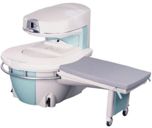
Manufactured by Esaote S.p.A.;
a low field open MRI scanner with permanent magnet for orthopedic use. The outstanding feature of this MRI system is a patient friendly design with 24 cm diameter, which allows the imaging of extremities and small body parts like shoulder MRI. The power consumption is around 1.3 kW and the needed minimum floor space is an area of 16 sq m.
At RSNA 2006 Hologic Inc. introduced a new dedicated extremity MRI scanner, the Opera. Manufactured by Esaote is the Opera a redesign of Esaote's 0.2 Tesla E-Scan XQ platform, which now enables complete imaging of all extremities, including hip and shoulder applications. 'Real-time positioning' reportedly speeds patient setup and reduces exam times.
Esaote North America and Hologic Inc are the U.S. distributors of this MRI device.
Device Information and Specification
CLINICAL APPLICATION
Dedicated extremity
CONFIGURATION
Extremity, shoulder (2), flex coil, knee dual phased array, ankle//foot dual phased array, hand//wrist dual phased array coil
PULSE SEQUENCES
SE, GE, IR, STIR, FSE, 3D CE, GE-STIR, 3D GE, ME, TME, HSE
IMAGING MODES
Single, multislice, volume study, fast scan, multi slab
TR
10 - 5000 msec
TE
6 - 110 msec
SINGLE SLICE
0.6 sec/image
MULTISLICE
0.6 sec/image
17 cm
2D: 2 mm - 10 mm;
3D: 0.6 mm - 10 mm
MEASURING MATRIX
256 x 256 maximum
PIXEL INTENSITY
4096 gray lvls, 256 lvls in 3D
MAGNET TYPE
Permanent
24 cm H, open
MAGNET WEIGHT
2250 kg, 4960 lbs
H*W*D
79 x 65 x 85 cm
POWER REQUIREMENTS
2,0 kW; 110/220 V single phase
STRENGTH
20 mT/m
5-GAUSS FRINGE FIELD, radial/axial
150 cm/130 cm
Passive
spacer

• View the DATABASE results for 'Opera (E-SCAN™ XQ)' (2).Open this link in a new window

 
Further Reading:
  News & More:
E-Scan, 510(k) Summary(.pdf)
Saturday, 15 May 2004   by www.accessdata.fda.gov    
Searchterm 'Phase' was also found in the following services: 
spacer
Radiology  (17) Open this link in a new windowUltrasound  (77) Open this link in a new window
Partial Averaging
 
Partial averaging is a scan time reduction method that takes advantage of the complex conjugate of the k-space. The number of phase encoding steps of the acquisition matrix are reduced in the phase encoding direction.
Since negative values of phase encoded measurements are identical to corresponding positive values, only a little over half (more than 62.5%) of a scan actually needs to be acquired to replicate an entire scan. This results in a reduction in scan time at the expense of signal to noise ratio. The time reduction can be nearly a factor of two, but full resolution is maintained.
Partial Fourier averaging can be used when scan times are long, the signal to noise ratio is not critical and where full spatial resolution is required. Partial averaging is particularly appropriate for scans with a large field of view and relatively thick slices; and in 3D scans with many slices. In some fast scanning techniques the use of partial averaging enables a shorter TE thus improving contrast.
Partial averaging is also called Fractional NEX, Half Scan, Half Fourier, Phase Conjugate Symmetry, Single Side Encoding.
spacer

• View the DATABASE results for 'Partial Averaging' (4).Open this link in a new window

MRI Resources 
Open Directory Project - Collections - Portals - Databases - Image Quality - Shoulder MRI
 
previous      56 - 60 (of 286)     next
Result Pages : [1 2 3 4 5 6 7]  [8 9 10 11 12 13 14 15 16 17 18 19 20 ... ]
 Random Page
 
Share This Page
FacebookTwitterLinkedIn

MR-TIP    
Community   
User
Pass
Forgot your UserID/Password ?    


Next big thing in MRI will be :
AI 
remote operator 
personalized protocols 
helium-free 
molecular MRI 
portable MRI 

Look
      Ups


MR-TIP.com uses cookies! By browsing MR-TIP.com, you agree to our use of cookies.

Magnetic Resonance - Technology Information Portal
Member of SoftWays' Medical Imaging Group - MR-TIP • Radiology-TIP • Medical-Ultrasound-Imaging • 
Copyright © 2003 - 2024 SoftWays. All rights reserved. [ 4 May 2024]
Terms of Use | Privacy Policy | Advertising
 [last update: 2024-02-26 03:41:00]