 | Info
Sheets |
| | | | | | | | | | | | | | | | | | | | | | | | |
 | Out-
side |
| | | | |
|
| | | | | |  | Searchterm 'Phase' was also found in the following services: | | | | |
|  |  |
| |
|
Oversampling is the increase in data to avoid aliasing and wrap around artifacts. Aliasing is the incorrectly mapping of tissue signal from outside the FOV to a location inside the FOV. This is caused by the fact, that the acquired k-space frequency data is not sampled density enough.
Oversampling in frequency direction, done by increasing the sampling frequency, prevents this aliasing artifact. The proper frequency based on the sampling theorem (Shannon sampling theorem/Nyquist sampling theorem) must be at least twice the frequency of each frequency component in the incoming signal. All frequency components above this limit will be aliased to frequencies between zero and half of the sampling frequency and combined with the proper signal information, which creates the artifact.
Oversampling creates a larger field of view, more data needs to be stored and processed, but this is for modern MRI systems not a real problem. Oversampling in phase direction ( no phase wrap), to eliminate wrap around artifacts, by increasing the number of phase encoding steps, results in longer scan/processing times. | | | | | | | | | | |  Further Reading: Further Reading: | Basics:
|
|
| |
| | | Searchterm 'Phase' was also found in the following services: | | | | |
| | |
| |
|
Quadrature detection is used in magnetic resonance imaging as well as in Doppler ultrasound and is also called quadrature demodulation or phase quadrature technique.
With this phase sensitive demodulation technique the complex demodulated signal is separated into two components. One is called the real channel; the second part is called the imaginary channel and is located 90° away from the real channel. The signals from both channels are combined to produce a single set of quadrature detected real and imaginary spectra. In MRI, the parts of the demodulated MR signal are further processed by Fourier transformation analysis. All information on the MR signal components e.g. amplitude, phase, and frequency is given by this quadrature detection combined with Fourier analysis. | | | |
• View the DATABASE results for 'Quadrature Detection' (2).
| | | | |
| | | | | |
| |
|
| | | |
• View the DATABASE results for 'Quadrature Detector' (7).
| | | | |
| | | Searchterm 'Phase' was also found in the following services: | | | | |
| | |
| |
|
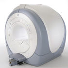
From GE Healthcare;
The Signa HDx MRI system is GE's leading edge whole body magnetic resonance scanner designed to support high resolution, high signal to noise ratio, and short scan times.
Signa HDx 3.0T offers new technologies like ultra-fast image reconstruction through the new XVRE recon engine, advancements in parallel imaging algorithms and the broadest range of premium applications. The HD applications, PROPELLER (high-quality brain imaging extremely resistant to motion artifacts), TRICKS (contrast-enhanced angiographic vascular lower leg imaging), VIBRANT (for breast MRI), LAVA (high resolution liver imaging with shorter breath holds and better organ coverage) and MR Echo (high-definition cardiac images in real time) offer unique capabilities.
Device Information and Specification CLINICAL APPLICATION Whole body
CONFIGURATION Compact short bore SE, IR, 2D/3D GRE, RF-spoiled GRE, 2DFGRE, 2DFSPGR, 3DFGRE, 3DFSPGR, 3DTOFGRE, 3DFSPGR, 2DFSE, 2DFSE-XL, 2DFSE-IR, T1-FLAIR, SSFSE, EPI, DW-EPI, BRAVO, Angiography: 2D/3D TOF, 2D/3D phase contrast vascular IMAGING MODES Single, multislice, volume study, fast scan, multi slab, cine, localizer H*W*D 240 x 2216,6 x 201,6 cm POWER REQUIREMENTS 480 or 380/415, 3 phase ||
COOLING SYSTEM TYPE Closed-loop water-cooled grad. | | | | | |
| | | Searchterm 'Phase' was also found in the following services: | | | | |
| | |
| |
|
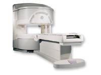
From GE Healthcare;
a friendly and less confining appearance targets the 7% of individuals who refuse to have an MRI because of claustrophobia. This open MRI system is also up to three times faster than other scanners, therefore the Signa OpenSpeed™ reducing exam time and scheduling
issues. In addition, a swing table provides better access and supports up to 500 pounds.
Device Information and Specification CLINICAL APPLICATION Whole body Standard: SE, IR, 2D/3D GRE and SPGR, Angiography: 2D/3D TOF, 2D/3D Phase Contrast;; 2D/3D FSE, 2D/3D FGRE and FSPGR, SSFP, FLAIR, EPI, optional: 2D/3D Fiesta, FGRET, Spiral, TensorTR 1.3 to 12000 msec in increments of 1 msec TE 0.4 to 2000 msec in increments of 1 msec 2D: 0.8mm - 20mm 3D: 0.1mm - 20mm 0.08 mm; 0.02 mm optional POWER REQUIREMENTS 200 - 480, 3-phase | | | |
• View the DATABASE results for 'Signa OpenSpeed™' (2).
| | | | | | Further Reading: | News & More:
|
|
| |
| | | | |
| | |
| | | |
|
| |
 | Look
Ups |
| |


