 | Info
Sheets |
| | | | | | | | | | | | | | | | | | | | | | | | |
 | Out-
side |
| | | | |
|
| | | | | |  | Searchterm 'Phase' was also found in the following services: | | | | |
|  |  |
| |
|
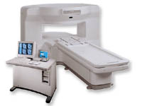
From GE Healthcare;
the New Signa Profile/i is a patient friendly open MRI system that virtually eliminates patient anxiety and claustrophobia, without compromising diagnostic utility.
Device Information and Specification CLINICAL APPLICATION Whole body Integrated transmit body coil, body flex sizes: M, L, XL, quadrature, head coil quadrature, 4 channel neurovascular array, 8 channel CTL array, quad. c- spine, 2 channel shoulder array, extremity coil, 3 channel wrist array, 4 channel breast array, 6, 9, 11 inch general purpose loop coils Standard: SE, IR, 2D/3D GRE and SPGR, Angiography: 2D/3D TOF, 2D/3D phase contrast; 2D/3D FSE, 2D/3D FRFSE, FGRE and FSPGR, SSFP, FLAIR, EPI, optional: 2D/3D Fiesta, fat/water separation, T1 FLAIRIMAGING MODES Localizer, single slice, multislice, volume, fast, POMP, multi slab, cine, slice and frequency zip, extended dynamic range, tailored RF TR 6 to 12000 msec in increments of 1 msec TE 1.3 to 2000 msec in increments of 1 msec 2D: 2.7mm - 20mm 3D: 0.2mm - 5mm 0.08 mm; 0.02 mm optional 10,000 kg w/gradient enclosure POWER REQUIREMENTS 200 - 480, 3-phase COOLING SYSTEM TYPE None required | | | | | |
| | | Searchterm 'Phase' was also found in the following services: | | | | |
| | |
| |
|
The respiratory phase can be used to control imaging either by only acquiring the image data during a particular portion of the respiratory cycle (which increases image acquisition time) or by adjusting the sequence of image data collection according to the phase of the respiratory cycle in such a way as to minimize motion-induced artifacts in the reconstructed image. See also Respiratory Ordered Phase Encoding. | | | | | |
| | | | | |
| |
|
Quick Overview
Please note that there are different common names for this artifact.
DESCRIPTION
Edge ringing, syrinx-like stripe
A data truncation artifact may occur when the interface between high and low signal intensities is encountered in one imaging plane. The 2D-FT techniques transform the MR signal to spatial intensity image data with frequency and phase information encoding each axis in the plane of the scan. This artifact is found in both frequency and phase axes.
Artifactual ripples adjacent to edges in an image or sharp features in a spectrum, caused by omission of higher frequency terms in Fourier transformation, particularly with the use of zero filling to replace unsampled higher frequencies.
Complex shapes are specified by series of sine and cosine waves of various frequencies, phase and amplitude. Some shapes are more difficult to encode than others. The most difficult shapes to represent with Fourier series of terms are waveforms with instantaneous transitions, tissue discontinuities or edges. The low-frequency components of the series describe the overall shape of the step function. Higher frequency components are needed to describe the corners if the step function more accurately.
If not enough samples are taken, these areas cannot be accurately represented.
The truncation of the infinite data series results in a ringing artifact because of the inability to accurately approximate this tissue discontinuity with a shorter truncated data set. Therefore, the ringing that occurs at all tissue boundaries on MR is called truncation artifact.

Image Guidance
| | | |
• View the DATABASE results for 'Truncation Artifact' (2).
| | | | |  Further Reading: Further Reading: | News & More:
|
|
| |
| | | Searchterm 'Phase' was also found in the following services: | | | | |
| | |
| |
|
| | | |
• View the DATABASE results for '2 Dimensional Fourier Transformation Imaging' (2).
| | | | | | Further Reading: | Basics:
|
|
| |
| | | Searchterm 'Phase' was also found in the following services: | | | | |
| | |
| |
|
(3D MRA) The 3D angiography technique can be applied to focus on fast flowing (arterial) blood and to visualize small tortuous vessels. 3D TOF images are less sensitive to turbulent flow artifacts.
The advantage of this approach is that the signal, acquired from the entire
volume has an increased signal to noise ratio. Slices are defined by a second phase encoded axis, which divides the volume into 'partitions'.
3D TOF MRA is acquired with 3D FT slabs or multiple overlapping thin 3D FT slabs ( MOTSA) depending on the coverage required and the range of flow-velocities under examination.
Such 3D techniques can provide equal spatial resolution along all three axes, i.e. be 'isotropic', or the partition thickness can be greater or less than the in plane spatial resolution in which case can be said to be 'anisotropic'.
The circle of Willis, anatomy as well as its fast arterial flow, lends itself well to both 3D TOF and 2D or 3D phase contrast angiography. | | | | | |
• View the DATABASE results for '3 Dimensional Magnetic Resonance Angiography' (2).
| | | | | | Further Reading: | Basics:
|
|
| |
| | | | |
| | |
| | | |
|
| |
 | Look
Ups |
| |


