 | Info
Sheets |
| | | | | | | | | | | | | | | | | | | | | | | | |
 | Out-
side |
| | | | |
|
| | | | |
Result : Searchterm 'Siemens' found in 1 term [ ] and 22 definitions [ ] and 22 definitions [ ] ]
| | previous 21 - 23 (of 23) Result Pages : [1] [2 3 4 5] |  | |  | Searchterm 'Siemens' was also found in the following services: | | | | |
| |  |
| |
|
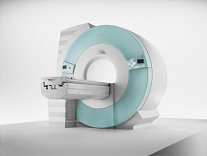
From Siemens Medical Systems;
70 cm + 125 cm + 1.5T and Tim - a combination never seen before in MRI ...
MAGNETOM Espree™s unique open bore design can accommodate more types of patients than other 1.5T systems on the market today, in particular the growing population of obese patients. The power of 1.5T combined with Tim technology boosts signal to noise, which is necessary to adequately image obese patients.
Device Information and Specification
CLINICAL APPLICATION
Whole body
Body, Tim [32 x 8], Tim [76 coil elements with up to 18 RF channels])
GRE, IR, FIR, STIR, TrueIR/FISP, FSE, FLAIR, MT, SS-FSE, MT-SE, MTC, MSE, EPI, 3D DESS//CISS/PSIF, GMR
IMAGING MODES
Single, multislice, volume study, multi angle, multi oblique
Image Processor reconstructing up to 3226 images per second (256 x 256, 25% recFoV)
1024 x 1024 full screen display
| | | | | | | | | | |  Further Reading: Further Reading: | News & More:
|
|
| |
| | | | | |
| |
|
•
In the 1930's, Isidor Isaac Rabi (Columbia University) succeeded in detecting and measuring single states of rotation of atoms and molecules, and in determining the mechanical and magnetic moments of the nuclei.
•
Felix Bloch (Stanford University) and Edward Purcell (Harvard University) developed instruments, which could measure the magnetic resonance in bulk material such as liquids and solids. (Both honored with the Nobel Prize for Physics in 1952.) [The birth of the NMR spectroscopy]
•
In the early 70's, Raymond Damadian (State University of New York) demonstrated with his NMR device, that there are different T1 relaxation times between normal and abnormal tissues of the same type, as well as between different types of normal tissues.
•
In 1973, Paul Lauterbur (State University of New York) described a new imaging technique that he termed Zeugmatography. By utilizing gradients in the magnetic field, this technique was able to produce a two-dimensional image (back-projection). (Through analysis of the characteristics of the emitted radio waves, their origin could be determined.) Peter Mansfield further developed the utilization of gradients in the magnetic field and the mathematically analysis of these signals for a more useful imaging technique. (Paul C Lauterbur and Peter Mansfield were awarded with the 2003 Nobel Prize in Medicine.)
•
1977/78: First images could be presented.
A cross section through a finger by Peter Mansfield and Andrew A. Maudsley.
Peter Mansfield also could present the first image through the abdomen.
•
In 1977, Raymond Damadian completed (after 7 years) the first MR scanner (Indomitable). In 1978, he founded the FONAR Corporation, which manufactured the first commercial MRI scanner in 1980. Fonar went public in 1981.
•
1981: Schering submitted a patent application for Gd-DTPA dimeglumine.
•
1982: The first 'magnetization-transfer' imaging by Robert N. Muller.
•
In 1983, Toshiba obtained approval from the Ministry of Health and Welfare in Japan for the first commercial MRI system.
•
1986: Jürgen Hennig, A. Nauerth, and Hartmut Friedburg (University of Freiburg) introduced RARE (rapid acquisition with relaxation enhancement) imaging. Axel Haase, Jens Frahm, Dieter Matthaei, Wolfgang Haenicke, and Dietmar K. Merboldt (Max-Planck-Institute, Göttingen) developed the FLASH ( fast low angle shot) sequence.
•
1988: Schering's MAGNEVIST gets its first approval by the FDA.
•
In 1991, fMRI was developed independently by the University of Minnesota's Center for Magnetic Resonance Research (CMRR) and Massachusetts General Hospital's (MGH) MR Center.
•
From 1992 to 1997 Fonar was paid for the infringement of it's patents from 'nearly every one of its competitors in the MRI industry including giant multi-nationals as Toshiba, Siemens, Shimadzu, Philips and GE'.
| | | | | |
• View the DATABASE results for 'MRI History' (6).
| | |
• View the NEWS results for 'MRI History' (1).
| | | | | | Further Reading: | | Basics:
|
|
News & More:
| |
| |
| | | | | |
| |
|
Silicon Graphics workstations are used in products from many other industry-leading medical manufacturers including:
The CT and MR products from General Electric Medical Systems uses Silicon Graphics® O2 and OCTANE workstation.
From Siemens Medical Systems, Inc., the postprocessing CT and MR workstations use Silicon Graphics O2 workstations.
Toshiba America MRI Inc. uses Silicon Graphics O2 workstations in its MRI scanners.
Bruker uses Silicon Graphics O2 workstations in its MRI scanners. | | | | | |
| | | Searchterm 'Siemens' was also found in the following services: | | | | |
| |
| | |
| | | |
|
| |
 | Look
Ups |
| |


