| Info
Sheets |
| | | | | | | | | | | | | | | | | | | | | | | | |
 | Out-
side |
| | | | |
|
| | | | |
Result : Searchterm 'mra' found in 2 terms [ ] and 57 definitions [ ] and 57 definitions [ ] ]
| | previous 6 - 10 (of 59) nextResult Pages : [1] [2 3 4 5 6 7 8 9 10 11 12] |  | |  | Searchterm 'mra' was also found in the following services: | | | | |
| |  |
| |
|
(SENSE) A MRI technique for relevant scan time reduction. The spatial information related to the coils of a receiver array are utilized for reducing conventional Fourier encoding. In principle, SENSE can be applied to any imaging sequence and k-space trajectories. However, it is particularly feasible for Cartesian sampling schemes. In 2D Fourier imaging with common Cartesian sampling of k-space sensitivity encoding by means of a receiver array enables to reduce the number of Fourier encoding steps.
SENSE reconstruction without artifacts relies on accurate knowledge of the individual coil sensitivities. For sensitivity assessment, low-resolution, fully Fourier-encoded reference images are required, obtained with each array element and with a body coil.
The major negative point of parallel imaging techniques is that they diminish SNR in proportion to the numbers of reduction factors.
R is the factor by which the number of k-space samples is reduced. In standard Fourier imaging reducing the sampling density results in the reduction of the FOV, causing aliasing. In fact, SENSE reconstruction in the Cartesian case is efficiently performed by first creating one such aliased image for each array element using discrete Fourier transformation (DFT).
The next step then is to create a full-FOV image from the set of intermediate images. To achieve this one must undo the signal superposition underlying the fold-over effect. That is, for each pixel in the reduced FOV the signal contributions from a number of positions in the full FOV need to be separated. These positions form a Cartesian grid corresponding to the size of the reduced FOV.
The advantages are especially true for contrast-enhanced MR imaging such as
dynamic liver MRI (liver imaging) ,
3 dimensional magnetic resonance angiography (3D MRA), and magnetic resonance cholangiopancreaticography ( MRCP).
The excellent scan speed of SENSE allows for acquisition of two separate sets of hepatic MR images within the time regarded as the hepatic arterial-phase (double arterial-phase technique) as well as that of multidetector CT.
SENSE can also increase the time efficiency of spatial signal encoding in 3D MRA. With SENSE, even ultrafast (sub second) 4D MRA can be realized.
For MRCP acquisition, high-resolution 3D MRCP images can be constantly provided by SENSE. This is because SENSE resolves the presence of the severe motion artifacts due to longer acquisition time. Longer acquisition time, which results in diminishing image quality, is the greatest problem for 3D MRCP imaging.
In addition, SENSE reduces the train of gradient echoes in combination with a faster k-space traversal per unit time, thereby dramatically improving the image quality of single shot echo planar imaging (i.e. T2 weighted, diffusion weighted imaging). | | | | | | | | | | |  Further Reading: Further Reading: | News & More:
|
|
| |
| | | | | |
| |
|
(3D MRA) The 3D angiography technique can be applied to focus on fast flowing (arterial) blood and to visualize small tortuous vessels. 3D TOF images are less sensitive to turbulent flow artifacts.
The advantage of this approach is that the signal, acquired from the entire
volume has an increased signal to noise ratio. Slices are defined by a second phase encoded axis, which divides the volume into 'partitions'.
3D TOF MRA is acquired with 3D FT slabs or multiple overlapping thin 3D FT slabs ( MOTSA) depending on the coverage required and the range of flow-velocities under examination.
Such 3D techniques can provide equal spatial resolution along all three axes, i.e. be 'isotropic', or the partition thickness can be greater or less than the in plane spatial resolution in which case can be said to be 'anisotropic'.
The circle of Willis, anatomy as well as its fast arterial flow, lends itself well to both 3D TOF and 2D or 3D phase contrast angiography. | | | | | |
• View the DATABASE results for '3 Dimensional Magnetic Resonance Angiography' (2).
| | | | | | Further Reading: | Basics:
|
|
| |
| | | | | |
| |
|
| | | |
• View the DATABASE results for 'Code 7228' (4).
| | | | | | Further Reading: | Basics:
|
|
| |
| | | Searchterm 'mra' was also found in the following services: | | | | |
| | |
| |
|
Contrast enhanced MRI is a commonly used procedure in magnetic resonance imaging. The need to more accurately characterize different types of lesions and to detect all malignant lesions is the main reason for the use of intravenous contrast agents.
Some methods are available to improve the contrast of different tissues. The focus of dynamic contrast enhanced MRI (DCE-MRI) is on contrast kinetics with demands for spatial resolution dependent on the application. DCE- MR imaging is used for diagnosis of cancer (see also liver imaging, abdominal imaging, breast MRI, dynamic scanning) as well as for diagnosis of cardiac infarction (see perfusion imaging, cardiac MRI). Quantitative DCE-MRI requires special data acquisition techniques and analysis software.
Contrast enhanced magnetic resonance angiography (CE- MRA) allows the visualization of vessels and the temporal resolution provides a separation of arteries and veins. These methods share the need for acquisition methods with high temporal and spatial resolution.
Double contrast administration (combined contrast enhanced (CCE) MRI) uses two contrast agents with complementary mechanisms e.g., superparamagnetic iron oxide to darken the background liver and gadolinium to brighten the vessels. A variety of different categories of contrast agents are currently available for clinical use.
Reasons for the use of contrast agents in MRI scans are:
•
Relaxation characteristics of normal and pathologic tissues are not always different enough to produce obvious differences in signal intensity.
•
Pathology that is sometimes occult on unenhanced images becomes obvious in the presence of contrast.
•
Enhancement significantly increases MRI sensitivity.
•
In addition to improving delineation between normal and abnormal tissues, the pattern of contrast enhancement can improve diagnostic specificity by facilitating characterization of the lesion(s) in question.
•
Contrast can yield physiologic and functional information in addition to lesion delineation.
Common Indications:
Brain MRI : Preoperative/pretreatment evaluation and postoperative evaluation of brain tumor therapy, CNS infections, noninfectious inflammatory disease and meningeal disease.
Spine MRI : Infection/inflammatory disease, primary tumors, drop metastases, initial evaluation of syrinx, postoperative evaluation of the lumbar spine: disk vs. scar.
Breast MRI : Detection of breast cancer in case of dense breasts, implants, malignant lymph nodes, or scarring after treatment for breast cancer, diagnosis of a suspicious breast lesion in order to avoid biopsy.
For Ultrasound Imaging (USI) see Contrast Enhanced Ultrasound at Medical-Ultrasound-Imaging.com.
See also Blood Pool Agents, Myocardial Late Enhancement, Cardiovascular Imaging, Contrast Enhanced MR Venography, Contrast Resolution, Dynamic Scanning, Lung Imaging, Hepatobiliary Contrast Agents, Contrast Medium and MRI Guided Biopsy. | | | | | | | | | | |
• View the DATABASE results for 'Contrast Enhanced MRI' (14).
| | |
• View the NEWS results for 'Contrast Enhanced MRI' (8).
| | | | | | Further Reading: | | Basics:
|
|
News & More:
|  |
FDA Approves Gadopiclenol for Contrast-Enhanced Magnetic Resonance Imaging
Tuesday, 27 September 2022 by www.pharmacytimes.com | | |
Effect of gadolinium-based contrast agent on breast diffusion-tensor imaging
Thursday, 6 August 2020 by www.eurekalert.org | | |
Artificial Intelligence Processes Provide Solutions to Gadolinium Retention Concerns
Thursday, 30 January 2020 by www.itnonline.com | | |
Accuracy of Unenhanced MRI in the Detection of New Brain Lesions in Multiple Sclerosis
Tuesday, 12 March 2019 by pubs.rsna.org | | |
The Effects of Breathing Motion on DCE-MRI Images: Phantom Studies Simulating Respiratory Motion to Compare CAIPIRINHA-VIBE, Radial-VIBE, and Conventional VIBE
Tuesday, 7 February 2017 by www.kjronline.org | | |
Novel Imaging Technique Improves Prostate Cancer Detection
Tuesday, 6 January 2015 by health.ucsd.edu | | |
New oxygen-enhanced MRI scan 'helps identify most dangerous tumours'
Thursday, 10 December 2015 by www.dailymail.co.uk | | |
All-organic MRI Contrast Agent Tested In Mice
Monday, 24 September 2012 by cen.acs.org | | |
A groundbreaking new graphene-based MRI contrast agent
Friday, 8 June 2012 by www.nanowerk.com |
|
| |
| | | | | |
| |
|
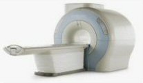
Device Information and Specification
CLINICAL APPLICATION
Whole body
SE, IR, FSE, FIR, GE, SG, BASG, PBSG, PCIR, DWI, Radial, Angiography: TOF, FLUTE (Fluoro-triggered bolus MRA), Time-resolved MRA
IMAGING MODES
Single, multislice, volume study
Level Range: -2,000 to +4,000
POWER REQUIREMENTS
208/220/240 V, single phase
| | | |
• View the DATABASE results for 'Echelon™ 1.5T' (2).
| | |
• View the NEWS results for 'Echelon™ 1.5T' (3).
| | | | | | Further Reading: | Basics:
|
|
| |
| | | | |
| | | |
|
| |
 | Look
Ups |
| |