| Info
Sheets |
| | | | | | | | | | | | | | | | | | | | | | | | |
 | Out-
side |
| | | | |
|
| | | | |
Result : Searchterm 'mra' found in 2 terms [ ] and 57 definitions [ ] and 57 definitions [ ] ]
| | previous 16 - 20 (of 59) nextResult Pages : [1] [2 3 4 5 6 7 8 9 10 11 12] |  | |  | Searchterm 'mra' was also found in the following services: | | | | |
| |  |
| |
|
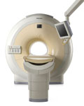
Device Information and Specification
CLINICAL APPLICATION
Whole body
CONFIGURATION
Short bore compact
Standard: Head, body, cardiac, optional phased array: Spine, pediatric, 3rd party connector; Optional SENSE? coils for all applications
SE, Modified-SE, IR (T1, T2, PD), STIR, FLAIR, SPIR, FFE, T1-FFE, T2-FFE, Balanced FFE, TFE, Balanced TFE, Dynamic, Keyhole, 3D, Multi Chunk 3D, Multi Stack 3D, K Space Shutter, MTC, TSE, Dual IR, DRIVE, EPI, Cine, 2DMSS, DAVE, Mixed Mode; Angiography: Inflow MRA, TONE, PCA, CE MRA
128 x 128, 256 x 256,512 x 512,1024 x 1024 (64 for Bold img)
Variable in 1% increments
Lum.: 120 cd/m2; contrast: 150:1
Variable (op. param. depend.)
POWER REQUIREMENTS
380/400 V
| | | | | |  Further Reading: Further Reading: | News & More:
|
|
| |
| | | | | |
| |
|
MS-325 is the formerly code name of gadofosveset trisodium (new trade name Vasovist). MS-325 belongs to a new class of blood pool agents for magnetic resonance angiography ( MRA) to diagnose vascular disease. Gadofosveset trisodium has ten times the signal-enhancing power of existing contrast agents as well as prolonged retention in the blood. This enables the rapid acquisition of high resolution MRA's using standard MRI machines.
Gadofosveset trisodium, which is gadolinium-based, stays in the blood stream as a result of transient binding to albumin. Albumin binding offers an additional benefit beyond localization in the blood pool. The contrast agent begins to spin much more slowly, at the rate albumin spins, causing a relaxivity gain that produces a substantially brighter signal than would be possible with freely circulating gadolinium.
MS-325 is an intravascular contrast agent intended for use in MRI as an aid in diagnosing aortoiliac occlusive disease in patients with known or suspected peripheral vascular disease (PVD) or abdominal aortic aneurysm (AAA).
Currently clinical trials completed for peripheral vascular disease and coronary artery disease. Additional trials are also being conducted to evaluate MS-325 as an aid in diagnosing breast cancer and suggested that it might be feasible to combine the use of MS-325, injected during peak stress, with delayed high-resolution imaging to identify myocardial perfusion defects.
Vasovist (MS-325) would compete with the contrast agents Ferumoxytol ( Code 7228) from AMAG Pharmaceuticals, Inc. and NC100150 Injection from Nycomed Amersham, but their further development is uncertain.
Partners in development: EPIX Pharmaceuticals, Inc., Mallinckrodt Inc., and Bayer Schering Pharma AG. Bayer Schering Pharma has the worldwide marketing rights for the product.
Formerly known under the Mallinckrodt trademark name, AngioMARK®.
See also Classifications, Characteristics, etc. | | | |
• View the DATABASE results for 'MS-325' (4).
| | |
• View the NEWS results for 'MS-325' (10).
| | | | | | Further Reading: | News & More:
|
|
| |
| | | | | |
| |
|
(PC) Phase contrast sequences are the basis of MRA techniques utilizing the change in the phase shifts of the flowing protons in the region of interest to create an image. Spins that are moving along the direction of a magnetic field gradient receive a phase shift proportional to their velocity.
In a phase contrast sequence two data sets with a different amount of flow sensitivity are acquired. This is usually accomplished by applying gradient pairs, which sequentially dephase and then rephase spins during the sequence. Both 2D and 3D acquisition techniques can be applied with phase contrast MRA.
The first data set is acquired with a flow compensated sequence, i. e. without flow sensitivity. The second data set is acquired with a flow sensitive sequence. The amount of flow sensitivity is controlled by the strength of the bipolar gradient pulse pair, which is incorporated into the sequence. Stationary tissue undergoes no effective phase change after the application of the two gradients. Caused by the different spatial localization of flowing blood to stationary tissue, it experiences a different size of the second bipolar gradient compared to the first. The result is a phase shift.
The raw data from the two data sets are subtracted. By comparing the phase of signals from each location in the two sequences the exact amount of motion induced phase change can be determined to have a map where pixel brightness is proportional to spatial velocity.
Phase contrast images represent the signal intensity of the velocity of spins at each point within the field of view. Regions that are stationary remain black while moving regions are represented as grey to white.
The phase shift is proportional to the spin's velocity, and this allows the quantitative assessment of flow velocities.
The difference MRI signal has a maximum value for opposite directions. This velocity is typically referred to as venc, and depends on the pulse amplitude and distance between the gradient pulse pair. For velocities larger than venc the difference signal is decreased constantly until it gets zero. Therefore, in a phase contrast angiography it is important to correctly set the venc of the sequence to the maximum flow velocity which is expected during the measurement. High venc factors of the PC angiogram (more than 40 cm/sec) will selectively image the arteries ( PCA - arteriography), whereas a venc factor of 20 cm/sec will perform the veins and sinuses (PCV or MRV - venography).
See also Flow Quantification, Contrast Enhanced MR Venography, Time of Flight Angiography, Time Resolved Imaging of Contrast Kinetics. | | | | | |
• View the DATABASE results for 'Phase Contrast Sequence' (5).
| | | | | | Further Reading: | Basics:
|
|
| |
| | | Searchterm 'mra' was also found in the following services: | | | | |
| | |
| |
|
| | | |
• View the DATABASE results for 'Time Resolved Imaging of Contrast Kinetics' (2).
| | | | | | Further Reading: | Basics:
|
|
| |
| | | | | |
| |
|
ABLAVAR™ (formerly named Vasovist™) is a blood pool agent for magnetic resonance angiography ( MRA), which opens new medical imaging possibilities in the evaluation of aortoiliac occlusive disease (AIOD) in patients with suspected peripheral vascular disease.
ABLAVAR™ binds reversibly to blood albumin, providing imaging with high spatial resolution up to 1 hour after injection, due to its high relaxivity and to the long lasting increased signal intensity of blood.
As with other contrast media: the possibility of serious or life-threatening anaphylactic or anaphylactoid reactions, including cardiovascular, respiratory and/or cutaneous manifestations, should always be considered.
WARNING:
NEPHROGENIC SYSTEMIC FIBROSIS
Gadolinium-based contrast agents increase the risk for nephrogenic systemic fibrosis (NSF) in patients with acute or chronic severe renal insufficiency (glomerular filtration rate less than 30 mL/min/1.73m 2), or acute renal insufficiency of any severity due to the hepato-renal syndrome or in the perioperative liver transplantation period.
See also Cardiovascular Imaging, Adverse Reaction, Molecular Imaging, and MRI Safety.
Drug Information and Specification
NAME OF COMPOUND
Diphenylcyclohexyl phosphodiester-Gd-DTPA, gadofosveset trisodium, MS-325
T1, predominantly positive enhancement
20-45 mmol-1sec-1, Bo=0,47T
PHARMACOKINETIC
Intravascular
CONCENTRATION
244 mg/mL, 0.25mmol/mL
DOSAGE
0.12 mL/kg, 0.03 mmol/kg
DEVELOPMENT STAGE
FDA approved
DO NOT RELY ON THE INFORMATION PROVIDED HERE, THEY ARE
NOT A SUBSTITUTE FOR THE ACCOMPANYING
PACKAGE INSERT!
Distribution Information
TERRITORY
TRADE NAME
DEVELOPMENT
STAGE
DISTRIBUTOR
USA, Canada, Australia
ABLAVAR™
Approved
| | | |
• View the DATABASE results for 'ABLAVAR™' (3).
| | |
• View the NEWS results for 'ABLAVAR™' (1).
| | | | | | Further Reading: | | Basics:
|
|
News & More:
| |
| |
| | | | |
| | |
| | | |
|
| |
 | Look
Ups |
| |